A 34 year old man already married came to us with erectile dysfunction.After basic evaluation he was started on PDE-5 inhibitors but the result was sub-optimal.
He was then given option of Intra-cavernosal Injection therapy with Bimix but he was repulsive for any injection over the penis.
His basic cause for erection was psychological so we though the medications along with Vacuum Erection Device would be good as he is bound to recover with thecourse of the time.
He opted for battery opted device and is happy with the usage.His wife has also accepted the method whole heartedly.
Vacuum erection devices, also known as vacuum constriction devices have been utilized for improving erectile rigidity for ovrigidity of the penile erection.
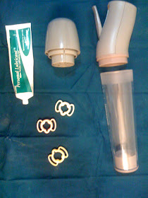
The vacuum device consists of a clear plastic cylinder with an aperture at one end that is placed over the penile shaft ; extending till the base of the penis. At the other end of the cylinder is a pump mechanism that is used to generate negative pressure within the cylinder. The pump mechanism can be in the form of either a manually operated(Figure 1 ) or a battery-operated system(Figure 2).
The former requires two hands to operate the device, one on the pump handle and the other to steady the cylinder on the penis itself. So this device is better to be given for people relatively yound and having dexterity over the movements.
The battery-operated device can be used with one hand. This is a better device in relatively infirm patients with neurological weaknesses.
Technique
Once a decision has been made to pursue sexual relations, water-soluble jelly is applied to the base of the penis (As shown in Figure). This maneuver helps in creating a water-tight seal, thus maintaining the negative pressure within the cylinder.
The patient can shave of the pubic hair for better fitting of the instrument. Once the cylinder has been placed over the penile shaft and held firmly against the pubic bone, the pump mechanism can be activated (either by hand held or battery). The negative pressure (vacuum generation) will cause blood to be drawn in the corpora cavernosa. The negative pressure build up is gradual and slow to prevent bruises and hematoma and resultant pain.
Once the erection is achieved the pressure can be let off and again rebuilt to maximize the erection . Once the erection has been achieved, a constriction ring (band) is applied to the base of the penis to act as an artificial valve, thus maintaining the blood within the corporal bodies. The rings come in a variety of shapes, sizes, and most importantly tension (tightness). The choice of the ring depends upon the patients penile size,turgidity and the patients preference.
Indications:
The vacuum device is indicated for men with ED. Especially in older patient and with cardiac comorbidities who are not suitable for medications.The patients who donot improve with the medications also can respond to the Vacuum Erection Device.
It is best suited for the patients with co-operative partner who accepts the usage of the Vacuum Erection Device.
Contra-indications:
(i) using antiplatelet agents/or presence bleeding disorders
(ii) history of priapism
(iii) congenital penile curvature/peyronies disease
(iv) psychiatric disorders/neurological disorders
There are instances in older people mistakenly the Vacuum Erection Device has been applied over the testis also along with the penis and resultant gangrene of the testis.It may so happen that in an inebriated patient may forget to remove the ring after the sexual act.The patient having neurological deficit and no sensations over the penis may neglect ongoing hematoma or even may forget to remove the ring causing grave implications.
It is generally advised to keep ring for not more than 30 minutes.It is essential in old couple to involve both the partners so that such problems can be avoided.
Important Facts to remember:
It has been estimated that the surface temperature of the penis during use of the VED is lower than the temperature prior to application of the device. The patient and his partner should be counseled regarding this fact prior to the initial use of the device as some couple might find cool penis repulsive for the sexual act. The device can take around 15 minutes to obtain erection.This may sometimes kill the already awakened sexual excitement /arousal .
Side Effects
1)cool penis sensations
2)Penile hematomas/bruising
3)The penile numbness might develop in some patients
4)The penis may loosely hand beyond the ring.
Satisfaction:
Despite the apparent drawbacks to the use of vacuum devices, there is a population of patients who find its use easy and it has allowed many couples to successfully resume penetrative sexual relations. The satisfaction rate varies from 35-80% but there are drop outs because of the side effects or some couple opting for better alternatives like penile implants which tries to imitate the natural erection.This device is not good for young people who may feel embarrassed to do whole action-applying the Vacuum Erection Device ;creating the pressure and carrying it everywhere as cumbersome.
Tuesday, May 25, 2010
Sunday, May 23, 2010
BOTOX: UTILITY IN UROLOGY
Botox, which has been smoothing wrinkles for years, now it will also help in relief of the bothersome urinary symptoms associated with an enlarged prostate or bladder conditions.
Botulinum toxin (BTX), a neurotoxin produced by the gram-positive, rod-shaped anaerobic bacterium Clostridium botulinum, was isolated in 1897 by Belgian scientist Professor Pierre Emile van Ermengem. BTX acts by blocking the release of acetylcholine at the neuromuscular junction. As a result of this chemodenervation, a temporary flaccid paralysis ensues. Different medical disciplines have taken advantage of this temporary paralysis to treat muscular hypercontraction. BTX was first approved by the US Food and Drug Administration in 1989 for use in patients with strabismus and blepharospasm. Since then, BTX has been used to treat a number of different neuromuscular disorders. BTX has been used successfully in urology to treat neurogenic and non-neurogenic detrusor overactivity, detrusor-sphincter dyssynergia, motor and sensory urge, and chronic pain syndromes.
The BOTOX toxin is of various types:A to F.The BOTOX A is the more potent with greater duration of action.It has wide urological applications.The BOTOX will bind irreversibly to presynaptic membrane and cause skeletal muscle atrophy but the axons will regenerate after 3-6 months.
BOTOX has been used in many urological consitions such as intractable overactive bladder,neurogenic bladder causing upper tract damage(kidney damage),Detrusor-External Sphicter Dyssenrgia( causing intermittent flow,obstructed stream in Neurological illnesses),chronic prostatic pain,non fibrotic bladder outflow obstruction(prostatic enlargement).
But the most common usage of BOTOX is in irritative bladder symptoms (frequency,nocturia,urgency,urge incontinence not yielding to medicines).The overactive bladder symptoms if doesnot abate with usual anti-cholinergic medications makes the life of the patient miserable. It will have physical problems-leakage causing personal inhygiene, psychological problems-embarrassment and loss of dignity, social problems-social isolation, sexual problems- because of genital skin rashes and foul smell( partner will have repulsion) etc.
The overactive bladder is widely prevalent affecting 50-100 millions of people all around the globe and some of them don’t respond to conventional treatments.These are the people who suffer silently and eventually end up in depression and self esteem.The BOTOX provides a ray of hope in such patients.
Typically 100-300 Units of BOTOX –A toxin is used. Briefly, the BoNT/A dose (200 or 300 units) is reconstituted with saline 0.9% at a total volume of 30 mL. The actual procedure of giving bladder Botox injections is fairly simple. It will take less than 20 minutes, and is minimally invasive. The procedure can be performed under local or general anaesthetic, and will not require an overnight stay in hospital.
A cystoscope – a small tube containing a camera – is passed into the bladder through the urethra so that the surgeon can inspect the inside of the bladder before performing the operation. A very thin needle is then passed through the cystoscope, and Botox is injected into between 20 and 30 different areas of the bladder muscle walls
BOTOX BEING INJECTED IN BLADDER FOR NEUROGENIC BLADDER
The BOTOX helps in alleviation of urological symptoms in 80% of the cases and the effect of BOTOX instillation lasts for 6-14 months.The injection can be repeated at those intervals.The BOTOX injection rarely causes systemic toxicity and rarely causes bladder paralysis needing long term catheterization.
One other area where BOTOX helps is neurogenic voiding dysfunction-either Detrusor Hyperreflexia or Detrusor External Sphincteric Dyssenergia in children with spina bifida/meningocele/myelomeningocele. These voiding dysfunction can gradually destroy the kidneys because of high bladder pressures.This can be brought down with BOTOX and kidney function thus preserved.
Certain novel areas like intractable chronic prostatitis and benign prostatic enlargement especially with detrusor overactivity ; many urologists have started using it with promising results.
The main implication of the BOTOX is that many patients having lower urinary tract symptoms with incontinence,urgency are elderly population with lot of other associated comorbidities like heart ailments.This makes them unsuitable for the conventional surgery if the medical line of treatment fails.In these group of patients BOTOX comes as a boon relieving them of the incontinence as it can be performed under local anaesthesia.
BOTOX although many people have reservations about its usage in urology is here to stay and its acceptance is going to increase because of its simplicity of performance and promising results.
Buccal Mucosal Graft Urethroplasty : A recent case
A -27- old gentleman came with history of weak stream, straining at micturition.There was no history of obvious trauma or prior urological intervention( like catheterization).He did not have the history of exposure also.There was no evidence of Balanitis Xerotica Obliternas on genital skin and mucosa.
He was evaluated and found to be short segment stricture in the proximal bulbar urethra.
He underwent multiple endoscopic interventions and urethral dilatations.
He needed recurrent dilatations. He was advised option of definitive urethroplasty.
He was taken up for ventral onlay urethroplasty.
Under spinal anesthesia through a midline perineal incision the bulbar urethra is exposed without mobilisation. Methylene Blue dye was injected through the meatus. A bougie was passed through the meatus upto the level of the stricture. Ventral urethrotomy is performed through the strictured urethra into normal proximal bulbar urethra upto 1.5cm. Methylene Blue stained urethral mucosa helps to identify the narrowed lumen of the urethra. A 2 cm wide and 6cm long buccal mucosal graft harvested from the cheek and it was defatted.
The BMG is sutured to the urethral mucosa with continuous sutures of 4/0 vicryl to the ventral urethrotomy throughout. A 14 F silastic Foley catheter was inserted to the bladder. The corpora spongiosa was over closed with continuous sutures of 4/0 vicryl and taking anchoring stitches through the buccal mucosa graft. The wound is closed in layers. The catheter is planned to be removed after 4 weeks.
Friday, May 21, 2010
Angioembolisation in Haemorrhagic cystitis
a 65 year old man presented to us with frank hematuria of 1 day duration. he was known case of small capacity bladder with hemorrhagic cystitis with no apparent reason.He was operated in 2002 for clam cystoplasty. He was apparently alright for 8 years just to land up in emergency department with gross total hematuria.
He is known case coronary artery disease and hypertension on medication.He was on ecosprin when he came for hematuria.
Immediately ecosprin was stopped.He was supported with irrigation,tranexa and cystoscopy and evacuation followed by alum irrigation.
After this surgery he was fine for 2 days then suddenly he had bout of frank hemturia causing fall of Hb from 13 TO 10 GM% and BP to fall from 130/80 mm Hg to 70/30 mmHg.
He was immediate taken up for cystoscopy and clot evacuation again with institution of proper blood support and plasma expander support.The bladder base region had angry looking globular mass? Rest of the bladder mucosa and the intestinal mucosa was normal.
The clots were removed with resectoscope and cautery and ellicke evacuator.A three way Foleys catheter was introduced wnd alum irrigation was started..The urine effluent was clear.
aFTER THE CLOT EVACUATION WAS DONE BILATERAL ANGIOEMBOLISATION WAS CARRIED OUT SELECTIVELY ON ANTERIOR DIVISION OF INTERNAL ILIAC ARTER USING SELDINGERS TECHNIQUE.The both iliac arteries anterior divison was blocked with gel foam mixture viscous with the contrast,
The process of Angioembolisation of the internal iliac artery --the end result of the embolisation is seen as the disapperance of the terminal branches of the vesical arteries.
The next plan was if the patient bleeds again then re-ileal conduit and extirpation of the diseased bladder at a later point of time.
The urological hemorrhage is an important problem in contemporary urological practice with significant associated morbidity and mortality. furthermore, these emergencies present a number of challenges to clinicians as current practice has evolved due to the increased availability of new imaging techniques and transarterial embolisation (tae). in this review we have explored the epidemiology, etiology and management of both renal and bladder hemorrhage. renal bleeding secondary to accidental or iatrogenic trauma and neoplastic disease requires careful but expeditious assessment and treatment. we have described current conservative, surgical and radiological approaches to the management of this challenging problem. moreover, bladder hemorrhage due to hemorrhagic cystitis, boadder cancer and infection represents a significant problem in current practice. advances in technology have changed the management options and again we have explored the literature in order to determine the optimum treatment approaches.
Wednesday, May 19, 2010
Post-Papavarine Injection Priapism:Management
A 35 year old gentleman presented with painful persistent penile erection after the injection of papavarine for penile doppler evaluation .The penile doppler and the intra-corporal injection were given 12 hours before.He had erection lasting for almost 12 hours before he presented to us.
The examination revealed tender turgid erecion with glans also rigid.
He was taken up for immediate intra-corporal aspiration with 21 G scalp vein.About 200 ml dark blood was evacuated followed which red blood started coming.
Then Phenyl Epinephrine (1 ml in 20 ml----500 mcg) was injected and kept for 5 moin.Then the rigidity was seen to subside.
But the tumuscence was still there.
One more dose was given and then the scalp vein was again clamped.Now this time the penis has become totally flaccid
PRIAPISM:
Priapism is erection that persists beyond or unrelated to sexual activity. It is of two types
Low flow-This is because of priapism due to lack of outflow leading to congestion of blood in corpora and subsequent decrease in arterial flow leading to ischemia. In this there is anoxia of smooth muscle component of corpora.
PRIAPISM IS AN EMERGENCY. Any delay in the treatement will result in corporal ischemia and fibrosis. This will lead to permanent erectile dysfunction and penile deformity.
High Flow: This is due to trauma to perineum causing arteriovenous fistula and increased flow. This is not an emergency.
History-Detailed history regarding Intracavernosal Injection of Vasoactive agents, Hematological diseases, substance abuse, perineal trauma should be taken. The duration and any accompanying pain should be inquired. Any past history of priapism should be inquired.
Clinical examination-
Very Important to feel for any Malignant induration (metastases causing priapism), Bruit in perineum (trauma related high flow priapism). These obviously pinpoint to aetiology and help in treatment.
Invstigations:
1) Duplex Doppler Ultrasound-to differentiate between low and high flow priapism. Duplex ultrasound will reveal low flow and constricted cavernosal artery while in high flow the flow will be turbulent indicative of arteriovenous fistula.
2) Cavernosal Blood gas Analysis (Important) Ph< 7.25, pCO2>60,pO2>30 (Low Flow).The arterial blood gas picture is reverse in High Flow variant. This is an important necessary tool because it definitively pinpoints the type of priapism. It involves aspiration of blood from the corpora and sending it to ABG analyser(usually present in ICU)
Management
Low flow variety
1) Hematological disorders-Always hydrate the patient first
2) If Priapism less than 4 hours-Intracavernosal Injection of Phenylephrine bolus 500mcg repeated after 5 minutes. Importantly patient’s vitals should be kept on monitoring.
3) If priapism more than 4 hours-Drain one corpora with 21 G scalp vein with aspiration to remove old anoxic blood and inject a bolus of Phenylephrine ( 1 ml of Phenylephrine with 19 ml Normal saline mixture).The scalp vein should be clamped for 5 minutes. Repeat the procedure if there is no response.
4) Alternatively drain the corpora with 20 G scalp vein passively and let the blood drain out on its own. Initially the drained blood is dark anoxic slowly once the smooth muscle component of the corpora recovers red blood oozes out and that is the end-point of the drainage.
5) No response-------Send to Andrologist for Performance of corpora-glanular shunt (AL GHORAB SHUNT) .This procedure is simple .It can be done under penile block anaesthesia. It involves peroration of the corpora cavernosa through spongiosum (glans).This is followed by dilatation of the fenestration by Hegar´s dilator. The aim is to allow drainage of anoxic cavernosal blood into relatively supple spongiosum. The glans wound is then closed .This procedure is safe and quick.
High flow variety:
Initially conservative treatment like application of pressure packing, ice packing, use of adrenergic agents as written above. If these measures fail then Internal pudental angiography and angioembolisation is the treatment.
Stuttering Priapism:
Many patients, especially children, have a pattern of multiple short episodes over a period of days or several weeks. The priapism is often normal flow and prognosis is generally good and therapy is conservative. If the episode lasts longer and turns painful; then it should be like low flow variety. The long term prevention can be done with Baclofen 40 mg at the bed time.
Tuesday, May 18, 2010
Andrology Department in Dr Ramayyas Hospital
We are glad to announce that a full fledged dedicated andrology department in Dr Ramayyas Urology Nephrology Hospital is functional.
We have started doing microsurgical reconstructive surgeries for male infertility and also for varicocele.
These are the facilities available in the hospital:
Semen analysis
Hormone profile measurement
Tran-rectal ultrasound
Testicular biopsy (diagnostic)
Vasography
Colour doppler ultrasound for the testes
Micro-surgical varicocelectomy
Microsurgical vasectomy reversals
Micro-surgical vaso-epididymostomy
Fertility preserving hernia and hydrocele repair
Trans-urethral ejaculatory duct resection
Seminal vesiculoscopy
No-scalpel vasectomy(NSV)
Sperm retrieval-microsurgical/open(Microdissection TESE)
Impotence evaluation and treatment
Medications for potency and pre- mature ejaculation
Bimix injections for potency
Vacuum erection devices/ penile rings
Penile venous ligation surgery
Penile implants
Penile revascularization surgery (post-trauma patients)
Surgery for penile curvatures and Peyronies disease
Monday, May 17, 2010
Urinary Incontinence:a Review
Stress Urinary Incontinence
1. An involuntary loss of urine during coughing, or physical exertion
2. Evident as leakage of urine on increased abdominal pressure without change in detrusor pressure (VLPP) during filling phase on UDS(SPECIALISED PRESSURE MANOMETRY)
It is a common and distressing problem, which may have a profound impact on quality of life. Urinary incontinence almost always results from an underlying treatable medical condition but is under-reported to medical practitioners.
Usual cause of stress urinary incontinence
1. Vaginal delivery--multiple vaginal births(unattended deliveries common in ESPECIALLY in villages)
2. Aging
3. Estrogen deficiency(Some woman leak one week before menstrual period.The lowered estrogen levels that particular time may lead to lower muscular pressure around the urethra, increasing chances of leakage. The incidence of stress incontinence increases following menopause, similarly because of lowered estrogen levels)
4. Neurological disease(especially diabetes)
5. In female high-level athletes, effort incontinence occurs in all sports involving abrupt repeated increases in intra-abdominal pressure that may exceed perineal floor resistance
As In India; multiple vaginal births are a common scenario and there is cultural taboo so incontinence is very high in prevalence but majority of household women suffer from it silently.Many of them avoid mingling in social occasions for fear of leakage preferring to remain aloof.The urine leakage is equally annoying to their sex partners which may severely affect sex life and adversely affect married life.There are certain myths in society about stress urinary leakage:
1. Urinary incontinence/prolapse is a natural part of aging
2. Nothing can be done about it
3. Surgery is the only solution(phobia for doctors;thinking that they will invariably suggest surgery for the disease)
Prevalence:
Reported prevalence rates range from 4.5% to 53%
Our Hopsital Statistics shows:
1. 50 Patients of stress/mixed incontinence / 6 months
2. 10 Undergo UDS/ 6months
3. 3-4 Undergo surgical intervention
Can we do something to remove doctor phobia especially in Indian society?
Can Nurse led continence service of any use?
A study was conducted by Matharu et al in 2004 where women aged ≥40 yrs with LUTS (n= 2421) were randomly allocated to a nurse-led continence service.Out of them , 450 underwent urodynamic study.The results showed women with OAB, 79.1% were correctly allocated anticholinergics & 64.8% were allocated pelvic floor training protocol(PFT).Of all women with urodynamic SUI, 88.8% were allocated PFT.This shows that nurse led continence service fairly treat women and this type of service can be initiated by Government of India to avoid urine leakage misery.
Management of tress urinary incontinence:
Continence and micturition involve a balance between urethral closure and detrusor muscle activity. Urethral pressure normally exceeds bladder pressure, resulting in urine remaining in the bladder. The proximal urethra and bladder are both within the pelvis. Intraabdominal pressure increases (from coughing and sneezing) are transmitted to both urethra and bladder equally, leaving the pressure differential unchanged, resulting in continence. Normal voiding is the result of changes in both of these pressure factors:urethral pressure falls and bladder pressure rises. SUI is due essentially to insufficient strength of the pelvic floor muscles. It is the loss of small amounts of urine associated with coughing, laughing, sneezing, exercising or other movements that increase intra-abdominal pressure and thus increase pressure on the bladder. The urethra is supported by fascia of the pelvic floor. If this support is insufficient, the urethra can move downward at times of increased abdominal pressure, allowing urine to pass.
So the basic aim of the treatment is Aim: To improve urethral resistance.These are the conservative measures:
1.Weight loss
A study published in The New England Journal of Medicine on January 29, 2009, demonstrated that weight loss in overweight women reduced stress incontinence. The study included women with a Body Mass Index (BMI) over 25 and at least 10 episodes of urinary incontinence per week. The results demonstrated that with exercise and restricted diet they had a 70% or greater reduction in overall incontinence episodes.So weigth loss should be the first thing a woman ahould follow for reduction in incontinence.
2. Absorbent products
Absorbent products include, undergarments, protective underwear, briefs, diapers and underpads.There are some assist devices used like vaginal pessaries,femsoft catheter as physical barrier for prevention of urinary leakage.
4. Exercises
One of the most common treatment recommendations includes exercising the muscles of the pelvis. Kegel exercises to strengthen or retrain pelvic floor muscles and sphincter muscles can reduce stress leakage.
Role of pelvic floor training:
The Cochrane Incontinence Group Specialized Trials Register included One arm comprised PFT, the other either no treatment, placebo, sham treatment. A total 13 trials involving 714 women were included.They concluded that PFT be included in first-line conservative management programs.Basically suffering woman should Identify the pubococcygeus muscle first with the help of urologist and then Exercise the muscle (10 s contraction followed by 10 s relaxation) 30 to 80 times /day.This Increases muscle support of the pelvic viscera &increased closing force on the urethra and the benefits may be seen in 2 to 6 weeks.
An alternative or adjunct to PME is exercises the pelvic muscles by holding small weights inside the vagina for up to 15 minutes bid.Successiely the weights can be increased I ncreasing the capacity of the pubococcygeus muscle contraction.Success rate up to 70% to 80%. A recent Cochrane Review shows no advantage to combining PFT with biofeedback over the use of well-done PFT alone.Atleast 3 months of pelvic floor exercises are necessary.
Biofeedback:
Biofeedback uses measuring devices to help the patient become aware of his or her body's functioning. By using electronic devices or diaries to track when the bladder and urethral muscles contract, the patient can gain control over these muscles. Biofeedback can be used with pelvic muscle exercises
RESULTS OF PELVIC FLOOR TRAINING:
There was a trial
1. 76 women underwent a 3-month exercise program & followed for 1 year.
2. 30% of subjects were cured &17% were improved.
3. Subjects with severe incontinence did not benefit from the therapy
Medications
Medications can reduce many types of leakage. Drugs with a-adrenergic activity to increase bladder outlet resistance.For Example ..phenylpropanolamine 25-75 mg bid &imipramine10-25 mg qd-tid.These medicines have been taken off from the U.S. market because of concerns about hemorrhagic strokes in young women.A new nedicine has been tried in SUI:duloxetene :called drug which kills three birds in one stone. It is Combined serotonin and nor-epinephrine re-uptake inhibitor.Its actions are :
o Increases tone of external urethral sphincter
o In an integrated analysis of 4 randomised controlled trials, it significantly decreased incontinence episode frequency by 51.5%
A study by Drutz et al revealed In a subgroup of women with severe SUI awaiting surgery, duloxetine was found to be effective.Incontinence decreased by 46% or their Incontinence Quality of Life (I-QOL) score improved by 6.3 points.
Vaginal oestrogens:
Vaginal oestrogens are used in SUI especially in aging population.The basis behind is
• Common embryonic origin of bladder urethra & vagina
• High concentration of estrogen receptors in pelvic tissues
• General collagen deficiency state (falconer et al., 1994)
• Urethral coaptation affected by loss of estrogen
The oestrogen cream can improve the mucosal integrity and suppleness of the urethra and the vagina thereby take care of the urethral coaptation.These medicines can produce harmful side effects if used for long periods. There is an increased risk of cancers of the breast and endometrium (lining of the uterus). A patient should talk to a doctor about the risks and benefits of long-term use of medications.
When should doctor send the patient for surgery?(Vague indicators)
1. Severe SUI(≥ 2 PADS /DAY)
2. Duration of symptoms> 5 years
3. VLPP≤80 cm H2O-Urdynamic parameters
Apart from that:
1. Pt with significant associated prolapse that may be corrected at the same time
2. High levels of physical stress owing to lifestyle or occupation-models,athletes,stage performers
Summary:
a) SUI needs to be treated with conservative measures initially: simple, inexpensive and without complications
b) No need of UDS prior to conservative measures
c) Duloxetine helpful in noncompliant pt.
Surgical Management of Stress Urinary Incontinence
Marshall Marchetti Krantz (MMK)
This procedure requires an abdominal incision. The bladder neck and urethra are separated from the back surface of the pubic bone. Sutures are placed on either side of the urethra and bladder neck, which are elevated to a higher position. The free ends of the stitches are anchored to surrounding cartilage and pubic bone.
Burch Colposuspension
This vaginal suspension procedure often is performed when the abdomen is open for another purpose, such as abdominal hysterectomy. The bladder neck and urethra are separated from the back surface of the pubic bone. The bladder neck then is elevated by lateral sutures that pass through the vagina and pubic ligaments.
Needle Suspension
Several needle suspension procedures have been developed, each named after its creator (e.g., Stamey, Raz, Gittes); however, the basic technique is the same. Essentially, sutures are placed through the pubic skin or a vaginal incision into the anchoring tissues on each side of the bladder neck and tied to the fibrous tissue or pubic bone.
Sling Procedures
Patients with severe stress incontinence and intrinsic sphincter deficiency may be candidates for a sling procedure. The goal of this treatment is to create sufficient urethral compression to achieve bladder control.
There are two techniques:
percutaneous, which requires a small abdominal incision, and
transvaginal, which is performed through the vagina.
Percutaneous slings
The pubovaginal sling is made of a strip of tissue from the patient's abdominal fascia (fibrous tissue). A synthetic sling may be used, but urethral tissue erosion commonly occurs.
An incision is made above the pubic bone, and a strip of abdominal fascia (the sling) is removed. Another incision is made in the vaginal wall, through which the sling is grasped and adjusted around the bladder neck. The sling is secured by two sutures loosely tied to each other above the pubic bone incision, providing a hammock to support the bladder neck.
Possible complications include accidental bladder injury, infection, and prolonged urinary retention, which may require chronic intermittent self-catheterization.
Transvaginal slings
No abdominal incision is required and a small incision is made in the vaginal wall. The permanenet tape is introduced via the vagina .The trocars are used to introduce the tape are removed through small incisions at both the sides of the inner thighs.
Overactive bladder:
• Overactive bladder (OAB) is a syndrome characterized by
Urgency
With or without urge incontinence
Usually accompanied by frequency and nocturia
Prevalence:
• Worldwide it is known to affect 50-100 million people
• OAB affects approximately 16%-22% of adult population.
• The Prevalence increases with advancing age.
National Overactive Bladder Evaluation (NOBLE) Study: Similar Prevalence Among Men and Women
Effects of overactive bladder:
1)Physical Problems:
Limitation of physical activities
Discomfort due to dampness
Unpleasant odour
Skin rashes/ ulcers
Confinement in nursing homes
Insomnia
Falls
2)Psychological problems:
Loss of independence — feels tied to home
Fear of embarrassment
Loss of dignity & self esteem
Affects career
Depression
Suicide
3)Social problems:
Reduction in social interaction/ increased social isolation
Alteration of travel plans (e.g. plan around availability of toilets)
Cessation of some hobbies
4) sexual problems:
Avoidance of sexual contact
Basic evaluation for patient of urgency
To be done in all patients
History & micturition diary
Physical examination
Laboratory tests
Supplementary assessments.
BUN, Serum creatinine.
Serum Glucose.
Urine cytology and AFB.
• Specialized tests must be tailored according to the questions that need to be answered:
Urodynamic Tests: in medication failure,prior to invasive therapy
Endoscopic tests:hematuria,sterile pyuria
Management of overactive bladder:
• Non-pharmacologic methods:
Bladder training/PFT: Pelvic Floor Muscle Training:
• Drawing in” or “lifting up” of peri-anal musculature with minimal contractions of abdomen, thigh and buttocks
• Contractions to be sustained for at least 10 seconds and done for 30-80 times/day for at least 8 weeks (Ferguson et al;1990)
• Standards for assessment of change in pelvic function not yet established
• Pharmacotherapy
Various drugs used in overactive bladder
• Botox:
Botox :A toxin –dose:300 U,
Injected intravesically(over 25-30 sites)
Causes afferent nerve denervation
Injection repeated every 6 monthly
• Neuromodulation:
S3 afferent nerve stimulation inhibits detrusor activity at the level of the sacral spinal cord
Sacral nerve stimulation therapy consists of two parts
An initial percutaneous nerve evaluation (PNE)
Followed by surgical implantation of a permanent electrode lead and pulse generator.
• Surgery:Augmentation cystoplasty:
Patch of detubularized intestine used to augment bladder
Complications (30-50%)
o Intestinal obstruction
o Need of CIC
o Calculus
o Metabolic complications
o Malignancy
1. An involuntary loss of urine during coughing, or physical exertion
2. Evident as leakage of urine on increased abdominal pressure without change in detrusor pressure (VLPP) during filling phase on UDS(SPECIALISED PRESSURE MANOMETRY)
It is a common and distressing problem, which may have a profound impact on quality of life. Urinary incontinence almost always results from an underlying treatable medical condition but is under-reported to medical practitioners.
Usual cause of stress urinary incontinence
1. Vaginal delivery--multiple vaginal births(unattended deliveries common in ESPECIALLY in villages)
2. Aging
3. Estrogen deficiency(Some woman leak one week before menstrual period.The lowered estrogen levels that particular time may lead to lower muscular pressure around the urethra, increasing chances of leakage. The incidence of stress incontinence increases following menopause, similarly because of lowered estrogen levels)
4. Neurological disease(especially diabetes)
5. In female high-level athletes, effort incontinence occurs in all sports involving abrupt repeated increases in intra-abdominal pressure that may exceed perineal floor resistance
As In India; multiple vaginal births are a common scenario and there is cultural taboo so incontinence is very high in prevalence but majority of household women suffer from it silently.Many of them avoid mingling in social occasions for fear of leakage preferring to remain aloof.The urine leakage is equally annoying to their sex partners which may severely affect sex life and adversely affect married life.There are certain myths in society about stress urinary leakage:
1. Urinary incontinence/prolapse is a natural part of aging
2. Nothing can be done about it
3. Surgery is the only solution(phobia for doctors;thinking that they will invariably suggest surgery for the disease)
Prevalence:
Reported prevalence rates range from 4.5% to 53%
Our Hopsital Statistics shows:
1. 50 Patients of stress/mixed incontinence / 6 months
2. 10 Undergo UDS/ 6months
3. 3-4 Undergo surgical intervention
Can we do something to remove doctor phobia especially in Indian society?
Can Nurse led continence service of any use?
A study was conducted by Matharu et al in 2004 where women aged ≥40 yrs with LUTS (n= 2421) were randomly allocated to a nurse-led continence service.Out of them , 450 underwent urodynamic study.The results showed women with OAB, 79.1% were correctly allocated anticholinergics & 64.8% were allocated pelvic floor training protocol(PFT).Of all women with urodynamic SUI, 88.8% were allocated PFT.This shows that nurse led continence service fairly treat women and this type of service can be initiated by Government of India to avoid urine leakage misery.
Management of tress urinary incontinence:
Continence and micturition involve a balance between urethral closure and detrusor muscle activity. Urethral pressure normally exceeds bladder pressure, resulting in urine remaining in the bladder. The proximal urethra and bladder are both within the pelvis. Intraabdominal pressure increases (from coughing and sneezing) are transmitted to both urethra and bladder equally, leaving the pressure differential unchanged, resulting in continence. Normal voiding is the result of changes in both of these pressure factors:urethral pressure falls and bladder pressure rises. SUI is due essentially to insufficient strength of the pelvic floor muscles. It is the loss of small amounts of urine associated with coughing, laughing, sneezing, exercising or other movements that increase intra-abdominal pressure and thus increase pressure on the bladder. The urethra is supported by fascia of the pelvic floor. If this support is insufficient, the urethra can move downward at times of increased abdominal pressure, allowing urine to pass.
So the basic aim of the treatment is Aim: To improve urethral resistance.These are the conservative measures:
1.Weight loss
A study published in The New England Journal of Medicine on January 29, 2009, demonstrated that weight loss in overweight women reduced stress incontinence. The study included women with a Body Mass Index (BMI) over 25 and at least 10 episodes of urinary incontinence per week. The results demonstrated that with exercise and restricted diet they had a 70% or greater reduction in overall incontinence episodes.So weigth loss should be the first thing a woman ahould follow for reduction in incontinence.
2. Absorbent products
Absorbent products include, undergarments, protective underwear, briefs, diapers and underpads.There are some assist devices used like vaginal pessaries,femsoft catheter as physical barrier for prevention of urinary leakage.
4. Exercises
One of the most common treatment recommendations includes exercising the muscles of the pelvis. Kegel exercises to strengthen or retrain pelvic floor muscles and sphincter muscles can reduce stress leakage.
Role of pelvic floor training:
The Cochrane Incontinence Group Specialized Trials Register included One arm comprised PFT, the other either no treatment, placebo, sham treatment. A total 13 trials involving 714 women were included.They concluded that PFT be included in first-line conservative management programs.Basically suffering woman should Identify the pubococcygeus muscle first with the help of urologist and then Exercise the muscle (10 s contraction followed by 10 s relaxation) 30 to 80 times /day.This Increases muscle support of the pelvic viscera &increased closing force on the urethra and the benefits may be seen in 2 to 6 weeks.
An alternative or adjunct to PME is exercises the pelvic muscles by holding small weights inside the vagina for up to 15 minutes bid.Successiely the weights can be increased I ncreasing the capacity of the pubococcygeus muscle contraction.Success rate up to 70% to 80%. A recent Cochrane Review shows no advantage to combining PFT with biofeedback over the use of well-done PFT alone.Atleast 3 months of pelvic floor exercises are necessary.
Biofeedback:
Biofeedback uses measuring devices to help the patient become aware of his or her body's functioning. By using electronic devices or diaries to track when the bladder and urethral muscles contract, the patient can gain control over these muscles. Biofeedback can be used with pelvic muscle exercises
RESULTS OF PELVIC FLOOR TRAINING:
There was a trial
1. 76 women underwent a 3-month exercise program & followed for 1 year.
2. 30% of subjects were cured &17% were improved.
3. Subjects with severe incontinence did not benefit from the therapy
Medications
Medications can reduce many types of leakage. Drugs with a-adrenergic activity to increase bladder outlet resistance.For Example ..phenylpropanolamine 25-75 mg bid &imipramine10-25 mg qd-tid.These medicines have been taken off from the U.S. market because of concerns about hemorrhagic strokes in young women.A new nedicine has been tried in SUI:duloxetene :called drug which kills three birds in one stone. It is Combined serotonin and nor-epinephrine re-uptake inhibitor.Its actions are :
o Increases tone of external urethral sphincter
o In an integrated analysis of 4 randomised controlled trials, it significantly decreased incontinence episode frequency by 51.5%
A study by Drutz et al revealed In a subgroup of women with severe SUI awaiting surgery, duloxetine was found to be effective.Incontinence decreased by 46% or their Incontinence Quality of Life (I-QOL) score improved by 6.3 points.
Vaginal oestrogens:
Vaginal oestrogens are used in SUI especially in aging population.The basis behind is
• Common embryonic origin of bladder urethra & vagina
• High concentration of estrogen receptors in pelvic tissues
• General collagen deficiency state (falconer et al., 1994)
• Urethral coaptation affected by loss of estrogen
The oestrogen cream can improve the mucosal integrity and suppleness of the urethra and the vagina thereby take care of the urethral coaptation.These medicines can produce harmful side effects if used for long periods. There is an increased risk of cancers of the breast and endometrium (lining of the uterus). A patient should talk to a doctor about the risks and benefits of long-term use of medications.
When should doctor send the patient for surgery?(Vague indicators)
1. Severe SUI(≥ 2 PADS /DAY)
2. Duration of symptoms> 5 years
3. VLPP≤80 cm H2O-Urdynamic parameters
Apart from that:
1. Pt with significant associated prolapse that may be corrected at the same time
2. High levels of physical stress owing to lifestyle or occupation-models,athletes,stage performers
Summary:
a) SUI needs to be treated with conservative measures initially: simple, inexpensive and without complications
b) No need of UDS prior to conservative measures
c) Duloxetine helpful in noncompliant pt.
Surgical Management of Stress Urinary Incontinence
Marshall Marchetti Krantz (MMK)
This procedure requires an abdominal incision. The bladder neck and urethra are separated from the back surface of the pubic bone. Sutures are placed on either side of the urethra and bladder neck, which are elevated to a higher position. The free ends of the stitches are anchored to surrounding cartilage and pubic bone.
Burch Colposuspension
This vaginal suspension procedure often is performed when the abdomen is open for another purpose, such as abdominal hysterectomy. The bladder neck and urethra are separated from the back surface of the pubic bone. The bladder neck then is elevated by lateral sutures that pass through the vagina and pubic ligaments.
Needle Suspension
Several needle suspension procedures have been developed, each named after its creator (e.g., Stamey, Raz, Gittes); however, the basic technique is the same. Essentially, sutures are placed through the pubic skin or a vaginal incision into the anchoring tissues on each side of the bladder neck and tied to the fibrous tissue or pubic bone.
Sling Procedures
Patients with severe stress incontinence and intrinsic sphincter deficiency may be candidates for a sling procedure. The goal of this treatment is to create sufficient urethral compression to achieve bladder control.
There are two techniques:
percutaneous, which requires a small abdominal incision, and
transvaginal, which is performed through the vagina.
Percutaneous slings
The pubovaginal sling is made of a strip of tissue from the patient's abdominal fascia (fibrous tissue). A synthetic sling may be used, but urethral tissue erosion commonly occurs.
An incision is made above the pubic bone, and a strip of abdominal fascia (the sling) is removed. Another incision is made in the vaginal wall, through which the sling is grasped and adjusted around the bladder neck. The sling is secured by two sutures loosely tied to each other above the pubic bone incision, providing a hammock to support the bladder neck.
Possible complications include accidental bladder injury, infection, and prolonged urinary retention, which may require chronic intermittent self-catheterization.
Transvaginal slings
No abdominal incision is required and a small incision is made in the vaginal wall. The permanenet tape is introduced via the vagina .The trocars are used to introduce the tape are removed through small incisions at both the sides of the inner thighs.
Overactive bladder:
• Overactive bladder (OAB) is a syndrome characterized by
Urgency
With or without urge incontinence
Usually accompanied by frequency and nocturia
Prevalence:
• Worldwide it is known to affect 50-100 million people
• OAB affects approximately 16%-22% of adult population.
• The Prevalence increases with advancing age.
National Overactive Bladder Evaluation (NOBLE) Study: Similar Prevalence Among Men and Women
Effects of overactive bladder:
1)Physical Problems:
Limitation of physical activities
Discomfort due to dampness
Unpleasant odour
Skin rashes/ ulcers
Confinement in nursing homes
Insomnia
Falls
2)Psychological problems:
Loss of independence — feels tied to home
Fear of embarrassment
Loss of dignity & self esteem
Affects career
Depression
Suicide
3)Social problems:
Reduction in social interaction/ increased social isolation
Alteration of travel plans (e.g. plan around availability of toilets)
Cessation of some hobbies
4) sexual problems:
Avoidance of sexual contact
Basic evaluation for patient of urgency
To be done in all patients
History & micturition diary
Physical examination
Laboratory tests
Supplementary assessments.
BUN, Serum creatinine.
Serum Glucose.
Urine cytology and AFB.
• Specialized tests must be tailored according to the questions that need to be answered:
Urodynamic Tests: in medication failure,prior to invasive therapy
Endoscopic tests:hematuria,sterile pyuria
Management of overactive bladder:
• Non-pharmacologic methods:
Bladder training/PFT: Pelvic Floor Muscle Training:
• Drawing in” or “lifting up” of peri-anal musculature with minimal contractions of abdomen, thigh and buttocks
• Contractions to be sustained for at least 10 seconds and done for 30-80 times/day for at least 8 weeks (Ferguson et al;1990)
• Standards for assessment of change in pelvic function not yet established
• Pharmacotherapy
Various drugs used in overactive bladder
• Botox:
Botox :A toxin –dose:300 U,
Injected intravesically(over 25-30 sites)
Causes afferent nerve denervation
Injection repeated every 6 monthly
• Neuromodulation:
S3 afferent nerve stimulation inhibits detrusor activity at the level of the sacral spinal cord
Sacral nerve stimulation therapy consists of two parts
An initial percutaneous nerve evaluation (PNE)
Followed by surgical implantation of a permanent electrode lead and pulse generator.
• Surgery:Augmentation cystoplasty:
Patch of detubularized intestine used to augment bladder
Complications (30-50%)
o Intestinal obstruction
o Need of CIC
o Calculus
o Metabolic complications
o Malignancy
Percutaneous Nephrostomy:Life saving measure in obstructed kidneys
A 50 year old lady came to us with history having tretaed for Carcinoma Cervix stage 3 B.She had recieved 4 fractions of EBRT outside.
She had history of pain in right flank and fever since 2 days.Her investigation revealed anemia,polymorphonucelocystosis,raised creatinine 11.4mg%
She was taken up for emergency PCN(percutanoeus nephrostomy).
The both pelvicalyceal systems were punctured with 20 G cheeba needle with ultrasound guidance.Then dyestudy was performed.
After the pelvicalyceal delineation,both the systems were punctured with 18 G PCN needle in posterio-inferior calyx.The guidewire was then placed and the tract was subsequently dilated till 16 fr.At the end of the procedure 14 Fr Malecots was placed in both systems.
There was hydronephrotic drip on both sides.She is supplemented with fluids and strict watch has been kept on her Electrolytes and ABG parameters to prevent post-obstructive diuresis and its complications.
Once the creatinine comes up she will be fit for stenting as the kink in ureter due to malignancy will disappear and she will be a good candidate for chemotherapy.
The facilities for emergency PCN are necessary at every Hospital and all the urologists should be well trained for the same as it can obviate the need for hemodialysis in such situations.It can be life saving measure and does not take more than 30 minutes.
She had history of pain in right flank and fever since 2 days.Her investigation revealed anemia,polymorphonucelocystosis,raised creatinine 11.4mg%
She was taken up for emergency PCN(percutanoeus nephrostomy).
The both pelvicalyceal systems were punctured with 20 G cheeba needle with ultrasound guidance.Then dyestudy was performed.
After the pelvicalyceal delineation,both the systems were punctured with 18 G PCN needle in posterio-inferior calyx.The guidewire was then placed and the tract was subsequently dilated till 16 fr.At the end of the procedure 14 Fr Malecots was placed in both systems.
There was hydronephrotic drip on both sides.She is supplemented with fluids and strict watch has been kept on her Electrolytes and ABG parameters to prevent post-obstructive diuresis and its complications.
Once the creatinine comes up she will be fit for stenting as the kink in ureter due to malignancy will disappear and she will be a good candidate for chemotherapy.
The facilities for emergency PCN are necessary at every Hospital and all the urologists should be well trained for the same as it can obviate the need for hemodialysis in such situations.It can be life saving measure and does not take more than 30 minutes.
Saturday, May 15, 2010
Renal cel Carcinoma:Review
The kidneys are dark-red, bean-shaped organs. There is a cavity attached to its concave side which drains into a tube which extends all the way to bladder.
Each Kidney is enclosed in a transparent membrane called the renal capsule which helps to protect them against infections and trauma. The kidney is divided into two main areas a light outer area called the renal cortex, and a darker inner area called the renal medulla. Within the medulla there are 8 or more cone-shaped sections known as renal pyramids. The areas between the pyramids are called renal columns.
Anatomy of the kidney:
What is renal cell carcinoma:
Renal cell carcinoma is the most common type of kidney cancer in adults. It occurs most often in men ages 50 - 70.
The exact cause is unknown.
Risk factors include:
• Dialysis treatment
• Family history of the disease/Genetics
• Horseshoe kidney
• Von Hippel-Lindau disease (a hereditary disease that affects the capillaries of the brain, eyes, and other body parts)
Renal cell carcimoma (RCC) is the third most common genitourinary cancer after prostate and bladder. Majority (80% to 85%) of kidney tumors are malignant. It is the most lethal malignancy of all urological cancers.
Unique characteristics of RCC
lack of early warning signs,
diverse clinical manifestations,
resistance to radiation and chemotherapy, and
immunogenic nature and spontaneous regressions.
What are the symptoms:
Now a days many renal cell carcinomas are detected incidentally during routine ultrasound examinations.
Otherwise the symptoms of renal cell carcinoma are:
Pain in flank (due to capsular distension)
Hematuria
Varicocele
Back pain
Systemic symptoms-fever,weight loss,loss of apettite
Anemia
Some times it can cause paraneoplastic symptoms like-hypertension,polycythemia
Diagnosis
Tests include:
• Abdominal CT scan: to see the size,extent and spread of the tumor
• Blood chemistry :Renal function tests
• Ultrasound of the abdomen and kidney : this is screening test
• Complete blood count (CBC): for anemia or polycythemia
• Intravenous pyelogram (IVP):Now –a days not routinely performed
• Liver function tests: staging work up
• Renal arteriography : sometimes necessary if Inferior Vena Cava spread then angioembolisation can downstage the tumor and make it less vascular and easier to operate.
• Urinalysis and urine cytology : sometimes Transitional Cell Carcinoma can mimick Renal cell Carcinoma which has different management after the initial surgery.
The following tests may be performed to see if the cancer has spread:
• Chest CT scan
• Bone scan
• MRI: especially if Inferior Vena Cava spread is suspected
• PET scan
Staging
Stage I
is an early stage of kidney cancer. The tumor measures up 7 centimeters
Stage II
is also an early stage of kidney cancer, but the tumor measures more 7 cm in size and the cancer is confined to the kidney.
Stage III is one of the following:
• The tumour has spread to adjacent renal vein or Inferior vena cava or lymph nodes
Stage IV is one of the following:
• The tumor extends beyond the fibrous tissue that surrounds the kidney(Gerotas Fascia);
• Cancer has distant spread
Treatment
Radical nephrectomy remains the treatment of choice for organ confined renal cell carcinoma.
The prototypical radical nephrectomy involves removal of the cancerous kidney outside the Gerotas fascia along with the adrenal and lymphadenectomy from aortic bifurcation to crus of diaphragm.(although there are a lot controversies about the lymphadenectomy)
This can be done by Laparoscopy which uses 4 -5 ports inside the abdomen and the organ is extracted with small lower abdominal incision.Sometimes morcellation can be used to avoid the incision.
In small tumour(less than 4 cm) especially in solitary kidney or multiple tumours both the kidney a partial removal of the kidney encompassing tumour with the 5-10 mm of the normal renal parenchyma is done either by open of laparoscopic method.
When the tumor has spread to Inferior vena cava- then extensive surgery with or without cardiac bypass is needed for complete extirpation of the malignancy.
In case of unfit patients or with multiple comorbidities or multiple tumours cryo-ablation of Radio-frequency ablation can be resorted to.
Laparoscopic Cryo-ablation in process
Advanced Renal Cell Carcinoma:
RCC diagnosed early can be managed with nephron sparing or radical nephrectomy with excellent 5 year survival and prognosis. The problematic cases are those presenting as advanced disease at the initial presentation. The advanced disease includes: T4 N0 M0, or any T, any N, M1. These cases are associated with poor survival and limited treatment options.
Options for chemotherapy and endocrine-based approaches are limited, and no hormonal or chemotherapeutic regimen is accepted as a standard of care. Therefore, various biologic therapies have been evaluated. New agents, such as sorafenib and sunitinib, having anti-angiogenic effects through targeting multiple receptor kinases, and have been investigated in patients failing immunotherapy.
Role of surgery
Palliative nephrectomy should be considered in patients with metastatic disease for alleviation of symptoms such as pain, hemorrhage, malaise. Several randomized studies are now showing improved overall survival in patients presenting with metastatic kidney cancer who have nephrectomy followed by either interferon or IL-2. If the patient has good physiological status, then nephrectomy should be performed prior to immunotherapy. There are anecdotal reports documenting regression of metastatic renal cell carcinoma after removal of the primary tumor but adjuvant nephrectomy is not recommended for inducing spontaneous regression; rather, it is performed to decrease symptoms or to decrease tumor burden for subsequent therapy in carefully controlled environments.
IMMUNOTHERAPY
The immune modulators, such as interferon, interleukins (IL-2, have been tried.
A : Interferons -The interferons are natural glycoproteins with antiviral, anti-proliferative, and immuno-modulatory properties. They have a direct anti-proliferative effect on renal tumor cells in vitro, stimulate host mononuclear cells, and enhance expression of major histocompatibility complex molecules.
Interferon-alpha, which is derived from leukocytes, has an objective response rate of approximately 15% (range 0-29%).
Interlukin -IL-2 is a T-cell growth factor and activator of T cells and natural killer cells. It hampers tumor growth by activating lymphoid cells in vivo without directly affecting tumor proliferation. It can be administered as high dose and low dose regimen.
A high-dose regimen (600,000-720,000 IU/kg q8h for a maximum of 14 doses) results in a 19% response rate with 5% complete responses. The majority of responses to IL-2 were durable, with median response duration of 20 months. Eighty percent of patients who responded completely to therapy with IL-2 were alive at 10 years. Most patients responded after the first cycle, and those who did not respond after the second cycle did not respond to any further treatment. Therefore, the current recommendation is to continue treatment with high-dose IL-2 to best response (up to 6 cycles) or until toxic effects become intolerable. Treatment should be discontinued after 2 cycles if the patient has had no regression.
Combinations of IL-2 and interferon or other chemotherapeutic agents such as 5-FU have not been shown to be more effective than high-dose IL-2 alone
Toxicity is dose dependent. Most common dose dependant toxicity is hypotension requiring vasopressors. Also malaise, diarrhea, pyrexia, and rashes are commonly reported toxicities.
Approved for treatment of patients with metastatic RCC.
Durability of response: Approximately 60% of CRs remain disease free at > 10 yrs follow-up
Biological Therapy:
Sunitinib (Sutent) Sunitinib is another multi-kinase inhibitor approved by the FDA in January 2006 for the treatment of metastatic kidney cancer that has progressed after a trial of immunotherapy. The approval was based on the high response rate (40% partial responses) and a median time to progression of 8.7 months and an overall survival of 16.4 months. The receptor tyrosine kinases inhibited by sunitinib include VEGFR 1-3 and PDGFR.
Major toxicities (grade II or higher) include fatigue (38%), diarrhea (24%), nausea (19%), dyspepsia (16%), stomatitis (19%), and decline in cardiac ejection fraction (11%).
A recent phase 3 study evaluating sunitinib in the first-line setting, compared against IFN-, in patients with metastatic RCC demonstrated significant improvement in PFS and response rates compared against the control arm. These results are considered to be preliminary, and longer-term follow-up is necessary for conclusive results.
Each Kidney is enclosed in a transparent membrane called the renal capsule which helps to protect them against infections and trauma. The kidney is divided into two main areas a light outer area called the renal cortex, and a darker inner area called the renal medulla. Within the medulla there are 8 or more cone-shaped sections known as renal pyramids. The areas between the pyramids are called renal columns.
Anatomy of the kidney:
What is renal cell carcinoma:
Renal cell carcinoma is the most common type of kidney cancer in adults. It occurs most often in men ages 50 - 70.
The exact cause is unknown.
Risk factors include:
• Dialysis treatment
• Family history of the disease/Genetics
• Horseshoe kidney
• Von Hippel-Lindau disease (a hereditary disease that affects the capillaries of the brain, eyes, and other body parts)
Renal cell carcimoma (RCC) is the third most common genitourinary cancer after prostate and bladder. Majority (80% to 85%) of kidney tumors are malignant. It is the most lethal malignancy of all urological cancers.
Unique characteristics of RCC
lack of early warning signs,
diverse clinical manifestations,
resistance to radiation and chemotherapy, and
immunogenic nature and spontaneous regressions.
What are the symptoms:
Now a days many renal cell carcinomas are detected incidentally during routine ultrasound examinations.
Otherwise the symptoms of renal cell carcinoma are:
Pain in flank (due to capsular distension)
Hematuria
Varicocele
Back pain
Systemic symptoms-fever,weight loss,loss of apettite
Anemia
Some times it can cause paraneoplastic symptoms like-hypertension,polycythemia
Diagnosis
Tests include:
• Abdominal CT scan: to see the size,extent and spread of the tumor
• Blood chemistry :Renal function tests
• Ultrasound of the abdomen and kidney : this is screening test
• Complete blood count (CBC): for anemia or polycythemia
• Intravenous pyelogram (IVP):Now –a days not routinely performed
• Liver function tests: staging work up
• Renal arteriography : sometimes necessary if Inferior Vena Cava spread then angioembolisation can downstage the tumor and make it less vascular and easier to operate.
• Urinalysis and urine cytology : sometimes Transitional Cell Carcinoma can mimick Renal cell Carcinoma which has different management after the initial surgery.
The following tests may be performed to see if the cancer has spread:
• Chest CT scan
• Bone scan
• MRI: especially if Inferior Vena Cava spread is suspected
• PET scan
Staging
Stage I
is an early stage of kidney cancer. The tumor measures up 7 centimeters
Stage II
is also an early stage of kidney cancer, but the tumor measures more 7 cm in size and the cancer is confined to the kidney.
Stage III is one of the following:
• The tumour has spread to adjacent renal vein or Inferior vena cava or lymph nodes
Stage IV is one of the following:
• The tumor extends beyond the fibrous tissue that surrounds the kidney(Gerotas Fascia);
• Cancer has distant spread
Treatment
Radical nephrectomy remains the treatment of choice for organ confined renal cell carcinoma.
The prototypical radical nephrectomy involves removal of the cancerous kidney outside the Gerotas fascia along with the adrenal and lymphadenectomy from aortic bifurcation to crus of diaphragm.(although there are a lot controversies about the lymphadenectomy)
This can be done by Laparoscopy which uses 4 -5 ports inside the abdomen and the organ is extracted with small lower abdominal incision.Sometimes morcellation can be used to avoid the incision.
In small tumour(less than 4 cm) especially in solitary kidney or multiple tumours both the kidney a partial removal of the kidney encompassing tumour with the 5-10 mm of the normal renal parenchyma is done either by open of laparoscopic method.
When the tumor has spread to Inferior vena cava- then extensive surgery with or without cardiac bypass is needed for complete extirpation of the malignancy.
In case of unfit patients or with multiple comorbidities or multiple tumours cryo-ablation of Radio-frequency ablation can be resorted to.
Laparoscopic Cryo-ablation in process
Advanced Renal Cell Carcinoma:
RCC diagnosed early can be managed with nephron sparing or radical nephrectomy with excellent 5 year survival and prognosis. The problematic cases are those presenting as advanced disease at the initial presentation. The advanced disease includes: T4 N0 M0, or any T, any N, M1. These cases are associated with poor survival and limited treatment options.
Options for chemotherapy and endocrine-based approaches are limited, and no hormonal or chemotherapeutic regimen is accepted as a standard of care. Therefore, various biologic therapies have been evaluated. New agents, such as sorafenib and sunitinib, having anti-angiogenic effects through targeting multiple receptor kinases, and have been investigated in patients failing immunotherapy.
Role of surgery
Palliative nephrectomy should be considered in patients with metastatic disease for alleviation of symptoms such as pain, hemorrhage, malaise. Several randomized studies are now showing improved overall survival in patients presenting with metastatic kidney cancer who have nephrectomy followed by either interferon or IL-2. If the patient has good physiological status, then nephrectomy should be performed prior to immunotherapy. There are anecdotal reports documenting regression of metastatic renal cell carcinoma after removal of the primary tumor but adjuvant nephrectomy is not recommended for inducing spontaneous regression; rather, it is performed to decrease symptoms or to decrease tumor burden for subsequent therapy in carefully controlled environments.
IMMUNOTHERAPY
The immune modulators, such as interferon, interleukins (IL-2, have been tried.
A : Interferons -The interferons are natural glycoproteins with antiviral, anti-proliferative, and immuno-modulatory properties. They have a direct anti-proliferative effect on renal tumor cells in vitro, stimulate host mononuclear cells, and enhance expression of major histocompatibility complex molecules.
Interferon-alpha, which is derived from leukocytes, has an objective response rate of approximately 15% (range 0-29%).
Interlukin -IL-2 is a T-cell growth factor and activator of T cells and natural killer cells. It hampers tumor growth by activating lymphoid cells in vivo without directly affecting tumor proliferation. It can be administered as high dose and low dose regimen.
A high-dose regimen (600,000-720,000 IU/kg q8h for a maximum of 14 doses) results in a 19% response rate with 5% complete responses. The majority of responses to IL-2 were durable, with median response duration of 20 months. Eighty percent of patients who responded completely to therapy with IL-2 were alive at 10 years. Most patients responded after the first cycle, and those who did not respond after the second cycle did not respond to any further treatment. Therefore, the current recommendation is to continue treatment with high-dose IL-2 to best response (up to 6 cycles) or until toxic effects become intolerable. Treatment should be discontinued after 2 cycles if the patient has had no regression.
Combinations of IL-2 and interferon or other chemotherapeutic agents such as 5-FU have not been shown to be more effective than high-dose IL-2 alone
Toxicity is dose dependent. Most common dose dependant toxicity is hypotension requiring vasopressors. Also malaise, diarrhea, pyrexia, and rashes are commonly reported toxicities.
Approved for treatment of patients with metastatic RCC.
Durability of response: Approximately 60% of CRs remain disease free at > 10 yrs follow-up
Biological Therapy:
Sunitinib (Sutent) Sunitinib is another multi-kinase inhibitor approved by the FDA in January 2006 for the treatment of metastatic kidney cancer that has progressed after a trial of immunotherapy. The approval was based on the high response rate (40% partial responses) and a median time to progression of 8.7 months and an overall survival of 16.4 months. The receptor tyrosine kinases inhibited by sunitinib include VEGFR 1-3 and PDGFR.
Major toxicities (grade II or higher) include fatigue (38%), diarrhea (24%), nausea (19%), dyspepsia (16%), stomatitis (19%), and decline in cardiac ejection fraction (11%).
A recent phase 3 study evaluating sunitinib in the first-line setting, compared against IFN-, in patients with metastatic RCC demonstrated significant improvement in PFS and response rates compared against the control arm. These results are considered to be preliminary, and longer-term follow-up is necessary for conclusive results.
Friday, May 14, 2010
ESWL:Non-Invasive treatment for stone fragmentaion
We had one 35 year old gentleman who presented to us with the bilateral renal calculi 1-1.5 cm with normal functioning kidneys.He was prestented in a view if the stones donot get fragmented with ESWL then after 4 weeks ,Retrograde Intra Renal Suregry (RIRS) can be done.
During the procedure there was good fragmentation.He is planned for review after 4 weeks and imaging at the same time.
Until 20 years ago, open surgery was necessary to remove a stone. The surgery required a recovery time of 4 to 6 weeks. Today, treatment for these stones is greatly improved, and many options do not require major open surgery and can be performed in an outpatient setting.
Extracorporeal Shock Wave Lithotripsy
ESWL or extracorporeal shock wave lithotripsy has revolutionized the treatment of renal stones. Kidney stones less than or equal to 1.5 cm in size in the kidney or upper ureter are best treated with ESWL.
Usually, this is an outpatient type of procedure using IV sedation or full anesthesia.
Treatment time runs from 1 to 2 hours. The stone is usually visualized with fluoroscopy and once centered for treatment, a shock wave is generated that penetrates the body and impacts upon the stone. After usually 3000 shocks are given, the stone gradually pulverizes, and the fragments are passed spontaneously over the next several days to weeks (It may sometimes take upto 3 months to pass all the fragments).
Complications of this procedure bleeding which is self resolving, infection so peri-procedure cover of antibiotics is essential. The third potential complication is sometimes the stone breaks and the gravels line up along the lower ureter making it necessary for the patient to undergo secondary/auxialiary procedure- Ureteroscopic clearance.
As with any procedure; pre-operative urine culture should be sterile and the patient should be off from the anti-platelet agents atleast for the 7 days for the safety otherwise large hematomas not only in kidneys but also in liver and adjacent organs are reported.
Thursday, May 13, 2010
Laparoscopic Cholecystectomy: Brief overview
Gall Bladder is a Pear shaped accessory digestive organ situated just beneath the liver.It is a small organ that aids digestion and stores bile produced by the liver(It can store uptom 50 ml of bile). In humans the loss of the gallbladder is not fatal... When the concentration of cholesterol or fats increases in the bile juice it precipitates as stone in the Gall Bladder. It can occur in all age groups and in both males and females, though more commonly in females. The five F’s of Gall Bladder stone diseases are “A Fat, Flatulent, Fair, Female of Forty is more likely to have gall stones”.
Laparoscopic cholecystectomy requires 3-4 small incisions in the abdomen to allow the insertion of operating ports, small cylindrical tubes approximately 5-10 mm in diameter, through which surgical instruments and a video camera are placed into the abdominal cavity(usually through the umbilical opening). The camera illuminates the surgical field and sends a magnified image from inside the body to a video monitor, giving the surgeon a close-up view of the organs and tissues. The surgeon watches the monitor and performs the operation by manipulating the surgical instruments through the operating ports.
To begin the operation, the patient is anesthetized and placed in the supine position on the operating table. A scalpel is used to make a small incision at the umbilicus. Using either a Veress needle(closed technique) or Hasson technique(open technique) the abdominal cavity is entered. The surgeon insufflates the abdominal cavity with carbon dioxide to create a working space.. Additional ports are placed inferior to the ribs at the epigastric, midclavicular, and anterior axillary positions. The gallbladder fundus is identified, grasped, and retracted superiorly. With a second grasper, the gallbladder infundibulum is retracted laterally to expose and open Calot's Triangle (the area bound by the cystic artery, cystic duct, and common hepatic duct). The triangle is gently dissected and then the cystic duct and the cystic artery are identified, clipped with tiny clips and cut. Then the gallbladder is dissected away from the liver bed and removed through one of the ports. This type of surgery requires meticulous surgical skill, but in straightforward cases can be done in about an hour.
Recently, this procedure is performed through a single incision in the patient's umbilicus. This advanced technique is called Laparoendoscopic Single Site Surgery or "LESS".
Wednesday, May 12, 2010
Androgen Deficiency in Aging Male and huge prostatomegaly: Dilemma of management
A patient presented to us in Ramayyas Urology and Nephrology Hospital with complaints of erectile dysfunction and lower urinary tract symptoms. As per the detailed history there was history of decrease libido, difficulty in maintaining the erection and frequent mood alterations also.
On examination the patient had bilateral hydrocele and normal testes(size and texture).The prostate showed Grade 4 enlargement .
The investigation revealed huge prostatomegaly (187 gms) and Sr PSA 11.81 NG/ML. His testosterone was subnormal 1.63 ng/ml( below the reference range).So the patient had grade 4 enlargement of prostate with high PSA and ADAMS(Androgen Deficiency of Aging Male )
Having tried Vacuum erection device which he felt was not suitable for his use and having not satisfied with the effect of PDE-5 inhibitors ;he was given option of testosterone supplementation.But taking into consideration high PSA there was chances of occult carcinoma which needed to be excluded.
The prostatic enlargement of 187 gm and high PSA with uroflowmetry showing Maximum Flow rate of 12 ml/sec also pointed out to benign prostatic hyperplasia which would need treatment (medically-Dutasteride would have complicated the erectile dysfunction and alpha blocker - there was chance of retrograde ejaculation).The surgical treatment was deemed to be optimum.
The all options were discussed with the patient.Finally a decision was made to go for PROSTATE BIOPSY(A 14 CORE BIOPSY), bladder neck sparing laser evaporation of the lateral lobe(pt had bilobar prostatomegaly-the right lobe was predominantly enlarged that was removed.This was done with two views: trying to preserve anterograde ejaculation and prevention further complication because of prostate enlargement after testosterone supplementation) and bilateral hydrocelectomy.
The prostatic biopsy report was negative for malignancy.He was started on Viagra( continuous therapy to take care of endothelial dysfunction rather as on when needed basis and testosterone supplementation once biopsy is negative) and androgen replacement therapy with Zandrova(testosterone cream 5 gm sachet locally) daily on shoulder after bath .
From andrological point of view he will be under complete follow-up with regular PSA and evaluation of improvement of erectile dysfunction.