Wednesday, March 31, 2010

Male Infertilty:Should we go for microsurgery wherever feasible rather than Assisted Reproductive Technology


There has been atleast 6 studies since 1973 which unambiguously show decrease in average sperm count as we are stepping into 21 st century.There has been estimated 5.2 million decrease in sperm count each year and the motilty has been reduced by 0.5% per year.This can be attributed to life style changes-smoking,substance abuse, increasing psychogenic stress,increasing prevalence of metabolic syndrome(obesity,hypertension),exposure to industrial/mutagenic pollutants and herbicides etc.This means more and more males will become infertiles and resort to medical/surgical or Assisted reproductive treatment.


Over the past 30 years,the treatment of infertility has seen the development of revolutionary new assisted reproduction technologies.First in 1978 Loius Brown - first baby to be concieved by In-Vitro Fertilisation and then it was micro-assisted Reproduction using techniques such as Intra-cytoplasmic sperm injection.


These highly complex technologies are used with increasing frequency in the treatment of couples around the globe;more than 1 million abies worldwide have been concieved by this manner.
These technologies bypass natural barriers so their is a risk of unwanted genetic traits being passed on to next generation .Researchers firmly believe that perhaps 75% of all infertility patients have a genetic basis.Then there is financial costs involved apart from mental agony if multiple cycles are required.
IVF and ICSI has been now routinely advised to all infertile males when medical management or Intra-Uterine Insemination fails.If evaluated properly the infertile males can be offered microsurgical treatment for allowing them to utilise a chance to parent their own biological children without or with a minimal aid of assisted reproduction.


Basically the infertile males can be grossly divided into two types of problems in sperm deficit in semen: Obstructive(defect in transport) or Non-Obstructive(defect in production).
In obstructive sperm problems, the bloackage can be treated in appropriate case by microsurgical reconstruction of the blocked passage(micro-surgical vaso-epididymostomy,microsurgical vasovasostomy).The chance of sperm reappearance rate can be as high as 80%.But unlike western populations; my observations about microsurgical success in India is less, probably because of the blockages here are more commonly due to infection.Infective blockages can be very difficult to treat even with microsurgery.
Microsurgery gives a magnification of 25-30 times.This magnification is highly necessary in reconstruction of vas/epididymis(male seminal pathway) as the naked eye cannot even visualise them properly so reconstructing them would be a remote possibility.


The advantage of microsurgical reconstruction is that once successful;natural conception is possible or minimal aid of Asssisted reproduction is necessary.I call it as one plus one free package as unlike IVF the couples donot have to resort to again same procedure for the second baby.
The multiple data have revealed cost effectiveness of microsurgical reconstruction over the routine sperm retrieval for IVF and ICSI.
For unobstructed oligospermia(problem with production); microsurgery has significant role in varicocecele(15-40% causative factor in male infertility).It can give success rate of almost 60%(pregnancy rate) in varicocele(although this issue is controversial- two randomised controlled trials have proven a role for surgery in varicocele related infertility).


One more area where it has a role is in some cases of male infertility that are advised donor insemination or adoption because of advanced testicular damage.In such cases microsurgical sperm retrieval(Micro-dissection TESE) can be utilised to find sperm in testis and this sperm can be used for Test Tube Baby process.The routine testicular sperm aspiration or biopsy may not yield sperms in grossly damaged testis but microsurgery can still offer a hope in such cases.


So I stress upon proper evaluation of male factor in infertility and offering them microsurgery in suitable cases before giving them an option for Assisted Reproduction Technology.

Tuesday, March 30, 2010

Pelvi-ureteric junction obstruction:overview

Ureteropelvic junction (UPJ) obstruction is defined as an obstruction of the flow of urine from the renal pelvis to the proximal ureter.


UPJ obstruction is the most common cause of neonatal and antenatal hydronephrosis, occurring in 1 per 1500 live births. 50% of patients diagnosed with antenatal hydronephrosis are eventually diagnosed with UPJ obstruction upon further workup.

PATHOPHYSIOLOGY:
Congenital UPJ obstruction most often results from intrinsic disease. A frequently found defect is the presence of an aperistaltic segment of the ureter, perhaps similar to that found in primary obstructive megaureter. In these cases, histopathologic studies reveal that the spiral musculature normally present has been replaced by abnormal longitudinal muscle bundles or fibrous tissue.
Other causes
A. An abnormal or high insertion of the ureter into the renal pelvis may affect drainage of urine. This may be an effect rather than a cause in some cases.
B. Crossing lower-pole renal vessel may result in pressure on the ureter by a vessel can prohibit urinary flow down the ureter.(cause and effect relationship not clear).
C. Rotation of the kidney and renal hypermobility can cause intermittent obstruction
D. Secondary UPJ obstruction can be caused by prior surgical intervention to treat other disorders (or failed repair of a primary UPJ obstruction (recurrent pelvi-ureteric junction obstruction). It is usually secondary to periureteral scar formation.


Problem
UPJ obstruction is defined as an obstruction of the flow of urine from the renal pelvis to the proximal ureter. The resultant back pressure within the renal pelvis may lead to progressive renal damage and deterioration.
UPJ obstruction presents most frequently in childhood, but adults and elderly individuals can also present
Frequency
• UPJ obstruction is found in approximately 50% of patients diagnosed with antenatal hydronephrosis.
• The male-to-female ratio of UPJ obstruction is 3-4:1.
• In general, the left kidney is more commonly affected than the right kidney.
• UPJ obstruction manifests bilaterally in 10% cases.
Presentation:

Neonates who present with hydronephrosis should be fully evaluated with voiding cystourethrography (VCUG; to rule out vesicoureteral reflux) and renal ultrasonography soon after birth.
If renal ultrasonography demonstrates hydronephrosis without reflux on VCUG, a diuretic renal scan should be performed to quantify relative renal function and to define the extent of obstruction.


Renal pelvic dilatation in a case of Pelvi-Ureteric Junction Obstruction


Micturating Cysto-Urethrography showing no reflux mandatory step before surgical therapy

Older children may present with UTI, a flank mass, or intermittent flank pain
Adults with UPJ obstruction can present with various symptoms, including back and flank pain, UTI, and/or pyelonephritis.
A detailed history may reveal that the pain correlates with periods of increased fluid intake or ingestion of a food with diuretic properties (ie, Dietl crisis, beer drinkers kidney).
Indications
The goals in treating patients with ureteropelvic junction (UPJ) obstruction are to improve renal drainage and to maintain or improve renal function.
As mentioned above, dilatation of the intrarenal collecting system or hydronephrosis does not necessarily imply obstruction. Specifically in children, renal pelvic dilatation should be monitored with serial imaging to assess for changes in dilatation, renal parenchymal thickness and/or the presence of scarring, and function(Renal ultrasonography and renal scintigraphy). Surgical repair is indicated upon a significant differential on serial imaging or progressive deterioration of renal function.
Similarly, in adults, repair is recommended if nuclear medicine renal scan(deteriorating on serial scan or deteriorated function on initial scan)or intravenous pyelography (IVP) reveals ureteral obstruction.
Relevant Anatomy

The evaluation of an obstructed ureteropelvic junction (UPJ) requires information about ureteral and surrounding anatomy, renal position and ectopy, associated vasculature, and renal function.
Prior to surgical intervention, the surgeon frequently evaluates for renal position/ectopy, mobility, and UPJ anatomy, such as high-insertion variants versus annular stricture variants.
The major vascular supply of the UPJ comes from branches of the renal artery. These vessels usually lie in an anteromedial location in relation to the proximal ureter. Aberrant polar vessels may also be associated with the renal pelvis, causing compression and obstruction of the collecting system. These vessels arise from either the renal artery from a position proximal to the main intrarenal branching site or directly from the aorta. They can surround the UPJ and can be associated with obstruction, or they may be aberrantly positioned secondary to increasing hydronephrosis.
The vascular anatomy at the UPJ becomes crucial during an endopyelotomy. The renal collecting system may be accessed percutaneously (antegrade) or in a retrograde fashion via passage of a ureteroscope through the urethra. While most associated UPJ vessels lie in the anteromedial plane, accessory vessels may lie posteriorly or laterally. If all endoscopic incisions are made in the posterior-lateral plane, intraoperative hemorrhage may occur. For this reason, a comprehensive vascular evaluation with complemented CT angiography is needed. CT scanning in combination with 3-phase and 3-dimensional contrast imaging yields a reported sensitivity of around 80% in revealing crossing vessels.


When an open or laparoscopic pyeloplasty is performed, an accurate understanding of the vascular anatomy allows the surgeon to preserve the accessory renal vessels and to redirect them if the surgeon feels that they contribute to the obstruction. If an endopyelotomy is planned, this information can guide the surgeon in directing the endopyelotomy incision away from crossing vessels.
Retrograde pyelography at the time of surgery is often used to estimate the length of the stricture and the amount of pelvis/ureter that needs to be excised at the time of the pyeloplasty to create a dependent funnel
Laboratory Studies
• All patients with possible ureteropelvic junction (UPJ) obstruction should be evaluated routine hematological and biochemical tests along with urinalysis.
Imaging Studies
• Renal ultrasonography and VCUG are performed in children with suspected UPJ obstruction.
• IVP is used to evaluate patients with possible UPJ obstruction. However, in the evaluation of a child with a hydronephrotic kidney, diuretic renography has taken the place of IVP. The benefits of diuretic renography are that iodine-based intravenous contrast is not used, radiation exposure is minimal, and renal function can be better quantified. The disadvantage of the nuclear medicine scan is that insight into renal anatomy is not obtained(especially many urologists are used to IVP rather than even CT reconstructed images for taking decisions regarding renal surgery for example-percutaneous nephrolithotomy).


IVP showing right pelvi-ureteric junction obstruction

• Functionally significant obstruction is often diagnosed with diuretic renal scanning. The conventional renographic criteria include a flat or rising washout curve after diuretic with T 1/2 of greater than 20 minutes and differential function of less than 40. The differential function is important in determining the need for intervention, especially in asymptomatic patients, and in selecting the appropriate treatment (pyeloplasty vs nephrectomy). Poorly functioning kidneys (<10%) are often best treated with nephrectomy. Nuclear medicine scanning is also used to assess outcomes after surgical intervention. The scans are best interpreted by combination of nuclear consultant and urologist as sometimes the sheer size of the pelvis and kidney can overestimate the function of the kidney. Also after the surgery situation may arise that the kidney size will decrease because of removal of excess of pelvis and removal of obstruction resulting in decreasing in relative function(not representative) so patients attendants and the patient should be counseled pre-operatively.


Renal scan showing obstructed kidney

• Sometimes in a patient the scan may come up with the equivocal findings in such situations the scan can be repeated with different protocoal(prior loading with diuretic so called F- protocol)

Medical Therapy

In children with ureteropelvic junction (UPJ) obstruction, GOAL is focused on maintaining sterile urine and assessing renal function and the degree of hydronephrosis. Typically, when imaging studies reveal an incomplete obstruction, the patient is monitored with routine renal ultrasonography and nuclear medicine renography.
Currently, medical therapy is unavailable for the treatment of both adult and pediatric cases of UPJ obstruction.
Initially, most children are treated conservatively and monitored closely. Intervention is indicated in the event of significantly impaired renal drainage or poor renal growth. The accepted criteria for intervention in infants and children include T 1/2 greater than 20 minutes, differential function less than 40%, and ongoing parenchymal thinning with or without contralateral compensatory hypertrophy(indicating the ipsilateral kidney atrophy) . Intervention is also indicated in those with pain, hypertension, hematuria, secondary renal calculi, and recurrent UTIs.( a word of caution –pain may not always be alleviated in renal surgery .The patient will need counseling regarding this prior)
Surgical Therapy

Surgical intervention to treat an obstructed UPJ is warranted, especially upon deterioration of renal function.
The principles of surgical repair, as initially described by Foley, include the following:
• Formation of a funnel
• Dependent drainage
• Watertight anastomosis
• Tension-free anastomosis


In children, the procedure of choice is an Anderson-Hynes dismembered pyeloplasty. The approach may be performed through a flank, dorsal lumbotomy, or anterior extraperitoneal technique. Laparoscopy has gained increasing acceptance in pediatric surgery and is often used to perform pyeloplasties in children. In many cases, laparoscopic pyeloplasty is technically unfeasible in very small children and infants because of space constraints. Using this method, the obstructed segment is completely resected, with reanastomosis of the renal pelvis and ureter in a dependent funneled fashion. The decision of whether to use a ureteral stent transiently during the initial healing process is based on the personal preference of the surgeon. The success rate of dismembered pyeloplasty for treating an obstructed UPJ exceeds 95%.

Open pyeloplasty leaves behind scar.Increased hospitalisation ,pain and delay in recuperation are the other problems.


Laparoscopic pyeloplasty offers a minimally invasive treatment option that may be used in patients with either primary or secondary UPJ obstruction and is emerging as a new criterion standard in the treatment of UPJ obstruction. Success rates are comparable with those of open pyeloplasty procedures, and some studies have shown that laparoscopy offers the advantages of decreased morbidity, shorter hospital stay, and quicker recovery. Laparoscopic pyeloplasty is a technically demanding procedure that generally requires significant laparoscopic experience. Robotic-assisted laparoscopic pyeloplasty has become increasingly popular as the robots have become more prevalent. A small intrarenal pelvis is a relative contraindication to laparoscopic pyeloplasty as the intrarenal dissection would pose difficulty for the laparoscopic surgeon.
Endoscopic treatment alternatives include an antegrade or retrograde endopyelotomy, which is an endoscopic incision performed through the obstructing segment.
Prior to incising a UPJ obstruction, imaging study(CT angiography) is recommended to evaluate adjacent ureteral vasculature.An endopyelotomy incision is performed through the area of obstruction with a laser, electrocautery, or endoscopic scalpel. Most surgeons dilate the newly incised area with a balloon catheter to help ensure a complete incision. This is followed by prolonged ureteral stenting, for a period of 4-8 weeks. The stent acts as internal scaffolding during healing and maintains renal drainage. Success rates with the percutaneous and ureteroscopic endopyelotomy are 80-90%.
When open pyeloplasty fails, endopyelotomy is particularly useful, even in the pediatric population.
In patients who have a suboptimal result from endopyelotomy, repeat incision can be performed with success. Traditional open or laparoscopic pyeloplasty is also indicated after failed endopyelotomy.
Of the open surgical repairs used to treat UPJ obstruction, the Anderson-Hynes dismembered pyeloplasty is particularly useful for the high-insertion variant. The benefit of this procedure is complete excision of the diseased segment of ureter and reconstruction with healthy viable tissue.

Spiral and vertical flaps (eg, Culp and DeWeerd, Scardino and Prince) are useful when a long-strictured segment of diseased ureter is encountered. With these procedures, the proximal ureter is re-created with redundant renal pelvis that is tubularized.
Ureterocalicostomy, ie, anastomosis of the ureter to a lower-pole renal calyx, is usually reserved for failed open pyeloplasty when no extrarenal pelvis and significant hilar scarring are present.It also is convenient to perform the procedure in thinned out parenchyma.
Robotic-assisted laparoscopic pyeloplasty


This procedure is particularly helpful in the surgeon who is learning the laparoscopic technique. The da Vinci robotic surgical system has been used successfully for laparoscopic reconstruction. It offers several advantages to surgeons unskilled in laparoscopy, including increased degrees of suturing freedom(just like movement of the wrist), stereoscopic vision, tremor filtration, precision and maneuvreability. The results are similar to those of conventional laparoscopic pyeloplasty.
Follow-up

Prophylactic antibiotic therapy should be given postoperatively. Remove the endopyelotomy stent after 4-8 weeks.
Follow up with renal ultrasonography 1-3 months after surgery. In addition, follow up with IVP or nuclear medicine renal scan 3-6 months after surgery.
Serial renal imaging is recommended for the first year after surgery and should be continued less frequently thereafter if results have normalized.
Complications

Potential complications from open surgical pyeloplasty include UTI and pyelonephritis, urinary extravasation and leakage, recurrent ureteropelvic junction (UPJ) obstruction, or stricture formation. Treatment of urinary leakage is centered around catheter drainage, such as nephrostomy, ureteral stent, or perianastomotic drain, to direct urine away from the perianastomotic tissues and to decrease the risk of postoperative stricture disease.
Specific complications from endopyelotomy include significant intraoperative bleeding if the endoscopic incision is made inadvertently into a major polar vessel, postoperative infection, and recurrence of obstruction. If significant intraoperative bleeding is encountered with hypotension, emergency arteriography and embolization are indicated.

Monday, March 29, 2010

Erectile dysfunction:Overview

“Man survives earthquakes, experiences the horrors of illness, and all of the tortures of the soul. But the most tormenting tragedy of all time is, and will be, the tragedy of the bedroom.”

Tolstoy


ED is the inability to achieve and maintain an erection adequate for intercourse to the mutual satisfaction of the man and his partner.
Prevalence:
The Massachusetts Male Aging Study - 52% of men between the ages of 40 and 70. (1987-1997).Erectile dysfunction is classified as minimal, moderate, or complete. Of the 52% of men who suffer from erectile dysfunction 17% have minimal ED, 25% have moderate ED, and 10% have complete ED.
The worldwide incidence of erectile dysfunction estimated at over 152 million men, with a forecast of 322 million men by the year 2025. This may be because of change in life style-increasing population suffering from metabolic syndrome(syndrome with increased cholesterol,hypertension,cardiac disease,hyperuricemia and diabetes),smoking and substance abuse.It may also be because of increasing stress and change in interpersonal relationships.

Pathophysiology:
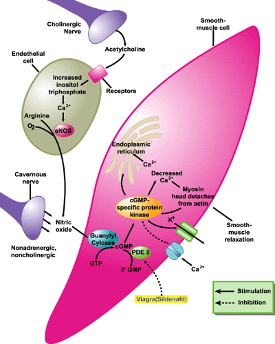
 The cGMP: The main mediator of penile erection causes smooth muscle relaxation and blood flows into the cavernous sinuses and the penis becomes erect.
 At the same time, the veins in the penis are squeezed almost completely shut due to this pressure. Since the veins are shut, blood can not drain from the penis, and it remains erect.
 Once the arousal has subsided, the cGMP is broken down and the penis again becomes flaccid.
 The main chemical responsible:phosphodiesterase-5 (PDE-5)sildenafil,Tadalafil are PDE-5 inhibitors :preventing the breakdown of cGMP, thus keeping blood in the penis to maintain the erection.


Causes
 Since an erection requires a precise sequence of events, ED can occur when any of the events is disrupted. The sequence includes nerve impulses in the brain, spinal column, and area around the penis, and response in muscles, fibrous tissues, veins, and arteries in and near the corpora cavernosa.

Damage to nerves, arteries, smooth muscles, and fibrous tissues, often as a result of disease, is the most common cause of ED. Erectile dysfunction is usually caused by one or more of the following pathologies::
 arterial vascular pathology: heart disease and vascular problems also raise the risk of erectile dysfunction.


 neurologic pathology
 Endocrine causes : Between 35 and 50 percent of men with diabetes experience ED. Also aging males can have dropping levels of testosterone which may lead to decreased libido,erectile dysfunction(Androgen Deficiency of Aging Male).Men with increased levels of prolactin will also experience erectile dysfunction because of decreased libido.
 Psychogenic causes Experts believe that psychological factors such as stress, anxiety(Performace anxiety), guilt(Previous sexual experiences), depression, low self-esteem, and fear of sexual failure cause 10 to 20 percent of ED cases.
 Post-Surgical: (especially radical prostate and bladder surgery for cancer) can injure nerves and arteries near the penis, causing ED.
 Local Pathology: Penile curvatures- Peyronies disease, corporal fibrosis-post trauma, surgery, post-priapism may also lead to impotence.
 Medications/substance abuse: many common medicines/drugs/substances-blood pressure drugs, antihistamines, antidepressants, tranquilizers, appetite suppressants,alcohol,tobacco and cimetidine (used for Acid Peptic Disease)-can produce ED


Evaluation:
 Somewhat subjective
 Doctor diagnosis
 Validated questionnaires like IIEF (International Index of Erectile Function)

Diagnosis
Patient History

Medical and sexual histories help define the degree and nature of ED. A medical history can disclose diseases that lead to ED, while a simple recounting of sexual activity might distinguish among problems with sexual desire,Painful sex,.dysfunction either difficulty in initiation or maintainance of erection, ejaculatory problems like-retrograde ejaculation or anejaculation, or problems with orgasm- anorgasmia.
Physical Examination
A physical examination can give clues to systemic problems. For example, if the penis is not sensitive to touching, Focal neurogical problems can pinpoint to neurological disorders.

• Abnormal secondary sex characteristics(defective androigenisation), such as hair pattern or breast enlargement, can point to hormonal problems, which would mean that the endocrine system is involved.
• The patient may have circulatory disturbances in the form of decreased pulsations in the ankle/popliteal region.
• Local pathologies: Penile curvatures, tender penile plaques will reveal local pathological cause for erectile dysfunction.

Practical Tips:
Psychogenic causes:

1. Younger patient (<40)
2. Preservation of morning erections and nocturnal erections
3. Achieve erection with masturbation
4. May be partner-specific(May achieve erection with specific person-quoad)
5. Often sudden onset(Related to sudden outburst of stress in life)
Organic ED:
1. Gradual deterioration over the period of months or years
2. Decrease in morning erections and nocturnal erections
3. No erections with masturbation
4. No loss of libido
5. Presence of co-morbid conditions like long standing hypertension,hyperlipidemia or diabetes.


Laboratory Tests

Several laboratory tests can help diagnose ED. Tests for systemic diseases include blood counts, urinalysis, lipid profile, and measurements of creatinine and liver enzymes. Measuring the amount of free and total testosterone, thyroid function or prolactin in the blood can yield information about problems with the endocrine system and is indicated especially in patients with decreased sexual desire.
Serum PSA is usually indicated in men over 50 years as if these men require testosterone replacement or supplementation they would need careful follow-up with Digital Rectal Examination and Serum PSA to rule out co-existing prostatic carcinoma which may flare up with the administration of testosterone.

Other Tests
Nocturnal Penile Tumuscence Tests:

Monitoring erections that occur during sleep (nocturnal penile tumescence) can help rule out certain psychological causes of ED. Healthy men have involuntary erections during sleep. If nocturnal erections do not occur, then ED is likely to have a physical rather than psychological cause.
In 1985, the RigiScan was introduced; it was the first device to provide automated, portable NPTR recording. The device combines the monitoring of radial rigidity, tumescence, number, and duration of erectile events with the convenience of a portable system that can be used at home. It consists of a recording unit that can collect data for three separate nights for a maximum of 10 hours each night. The mechanics consist of two loops: one is placed at the base of the penis and the other at the coronal sulcus. By constricting the loops, the device records penile tumescence (circumference) and radial rigidity at the penile base and tip. Measurement (initialization) is first done in the office for 15 to 20 minutes with the patient awake to establish an individual baseline. At home, penile rigidity is registered every 3 minutes by constriction of the loops, applying a radial compression.Radial rigidity above 70% represents a nonbuckling erection, and a rigidity of less than 40% represents a flaccid penis. The number of erections considered normal is three to six per 8-hour session, lasting an average of 10 to 15 minutes each
Cilurzo and colleagues (1992) recommend the following as normal NPTR criteria: four to five erectile episodes per night; mean duration longer than 30 minutes; an increase in circumference of more than 3 cm at the base and more than 2 cm at the tip; and maximal rigidity above 70% at both base and tip
The main advantages of NPT testing are its relative freedom from psychologic influences and its ability to detect sleep-related abnormalities. The documented presence of a full erection indicates that the neurovascular axis is functionally intact and that the cause of the ED is most likely psychogenic. Heaton and Morales (1997) have suggested indications for NPTR as follows: (1) suspected sleep disorder; (2) obscure cause of ED; (3) nonresponse to therapy; (4) planned surgical treatment; (5) legally sensitive case; (6) measurement of drug effects in placebo-controlled drug trials; and (7) suspected psychogenic cause.
Colour Doppler examination:
When vascular evaluation is indicated, intracavernous injection with color duplex Doppler ultrasound is the most informative diagnostic test. This may be all that is needed to define and determine severity. Color duplex ultrasound should be used before other tests are considered because it is the least invasive technology for evaluating vascular ED, distinguishing high-from low-flow priapism, and assessing Peyronie's plaque. Recently, the combination of oral sildenafil citrate with visual erotic stimulation has been shown to be an effective noninvasive pharmacologic induction method for penile blood flow evaluation rather than giving intracavernosal injection(which many patients find repulsive at first instance) PSV less than 25 cm/s after intracavernous injection and sexual stimulation has a 100% sensitivity and 95% specificity in selecting patients with abnormal penile arteriography, because it reflects severe cavernous arterial insufficiency. A PSV consistently greater than 35 cm/s is associated with normal arteriography and defines normal cavernous arterial inflow.
Penile arteriography:
Arteriography is most useful in providing anatomic information. The inferior epigastric vessels also need to be studied because they are most commonly used for penile revascularization. Because of the high cost and invasive nature of the study, only a small percentage of patients with complex ED are appropriate candidates- ED secondary to a traumatic arterial disruption or in a patient with a history of perineal compression injury. In these highly selected cases, a detailed “road map” of the arterial anatomy is essential to planning surgical reconstruction.
Dynamic Infusion Cavernosography and Cavernosometry:
This is a complex test done in special circumstances like young men who might be candidates for penile vascular operations, specifically those with a history of pelvic trauma.
Psychosocial Examination
A psychosocial examination, using an interview and a questionnaire, reveals psychological factors. A man's sexual partner may also be interviewed to determine expectations and perceptions during sexual intercourse.
Treatment
General Conservative measures:


Most physicians suggest that treatments proceed from least to most invasive. For some men, making a few healthy lifestyle changes may solve the problem. Quitting smoking, losing excess weight, and increasing physical activity may help some men regain sexual function. Avoiding drugs with harmful side effects is considered next. For example, drugs for high blood pressure like thiazides can be changed and alternative medications can be given.


Psychotherapy

Experts often treat psychologically based ED using techniques that decrease the anxiety associated with intercourse. The patient's partner can help with the techniques, which include gradual development of intimacy and stimulation.Master and Johnsons therapy involving couples can solve problems with performance anxiety /premature ejaculation
Drug Therapy
Drugs for treating ED can be taken orally, injected directly into the penis, or inserted into the urethra at the tip of the penis. In March 1998, the Food and Drug Administration (FDA) approved Viagra, the first pill to treat ED. Since that time, vardenafil hydrochloride (Levitra) and tadalafil (Cialis) have also been approved. Additional oral medicines are being tested for safety and effectiveness. Viagra, Levitra, and Cialis all belong to a class of drugs called phosphodiesterase (PDE) inhibitors. Taken an hour before sexual activity, these drugs work by enhancing the effects of nitric oxide, a chemical that relaxes smooth muscles in the penis during sexual stimulation and allows increased blood flow.

While oral medicines improve the response to sexual stimulation, they do not trigger an automatic erection as injections do. The recommended dose for Viagra is 50 mg, and the physician may adjust this dose to 100 mg or 25 mg, depending on the patient. The recommended dose for either Levitra or Cialis is 10 mg, and the physician may adjust this dose to 20 mg if 10 mg is insufficient. A lower dose of 5 mg is available for patients who take other medicines or have conditions that may decrease the body's ability to use the drug. Levitra is also available in a 2.5 mg dose.

None of these PDE inhibitors should be used more than once a day. Men who take nitrate-based drugs such as nitroglycerin for heart problems should not use either drug because the combination can cause a sudden drop in blood pressure. Also patients have severe cardiac diseases like recent Myocardial Infarction, reduced stress tolerance should avoid PDE-5 inhibitors.

Many men achieve stronger erections by injecting drugs into the penis, causing it to become engorged with blood. Drugs such as papaverine hydrochloride, phentolamine, and alprostadil (marketed as Caverject) widen blood vessels. These drugs may create unwanted side effects, however, including pain (36%) persistent erection (4%) and scarring. It gives a success rate of 70-90 % but these injections have a drop-out rate of 25-60% because of mainly pain or sometimes development of corporal fibrosis. A system for inserting a pellet of alprostadil into the urethra is marketed as Muse. The system uses a prefilled applicator to deliver the pellet about an inch deep into the urethra. An erection will begin within 8 to 10 minutes and may last 30 to 60 minutes. The most common side effects are aching in the penis, testicles, and area between the penis and rectum; warmth or burning sensation in the urethra; redness from increased blood flow to the penis; and minor urethral bleeding or spotting.


Vacuum Devices

Mechanical vacuum devices cause erection by creating a partial vacuum, which draws blood into the penis, engorging and expanding it. The devices have three components: a plastic cylinder, into which the penis is placed; a pump, which draws air out of the cylinder; and an elastic band, which is placed around the base of the penis to maintain the erection after the cylinder is removed and during intercourse by preventing blood from flowing back into the body The satisfaction rate varies from 35-80% but the problems with the Vacuum Erection devices are – cold penis, loosely hanging penis ,sometimes it can lead to bruises especially patient using it roughly and on aspirin or clopidogrel.


Surgery
Surgery usually has one of three goals:
•to implant a device that can cause the penis to become erect (Penile Implant surgery)
•to reconstruct arteries to increase flow of blood to the penis (Penile revascularization surgery for patient with focal arterial stenosis-post-trauma)
•to ligate veins that allow blood to leak from the penile tissues (penile venous leak-particularly detected on Doppler showing persistence end-diastolic velocity more than 5 cm/sec)

Implanted devices, known as prostheses, can restore erection in many men with ED. Possible problems with implants include mechanical breakdown and infection, although mechanical problems have diminished in recent years because of introduction of Viagra but there are a group of patients who fail with medications and refuse or fail with Vacuum Erection Device. Inflatable implants consist of paired cylinders, which are surgically inserted inside the penis and can be expanded using pressurized fluid Tubes connect the cylinders to a fluid reservoir and a pump, which are also surgically implanted. The patient inflates the cylinders by pressing on the small pump, located under the skin in the scrotum. Inflatable implants can expand the length and width of the penis. They also leave the penis in a more natural state when not inflated.


Advantages with the penile impants are:
 Good rigidity
 Freedom from medications
 Outpatient/24HR surgery
 Resume sexual activity 4-6 weeks
 No loss of ability to ejaculate or achieve orgasm


Surgery to repair arteries can reduce ED caused by obstructions that block the flow of blood. The best candidates for such surgery are young men with discrete blockage of an artery because of an injury to perineum or fracture of the pelvis.

Surgery to veins that allow blood to leave the penis usually involves an opposite procedure-intentional blockage. Blocking off veins (ligation) can reduce the leakage of blood that diminishes the rigidity of the penis during erection. However the results are not long lasting so the venous ligation surgery have diminished

Saturday, March 27, 2010

Seminal Vesiculoscopy with Ejaculatory Duct Obstruction

A 39 year old gentleman came with low volume ejaculate with oligospermia(documented on several semen analyseserum total tests) The semen volume was characteristically around 0.25-0.30 ml.His post-ejaculatory urine analysis for sperms was negative.His libido and orgasm was normal. He was evaluated with Hormone Profile(LH,FSH and Testosterone),Trans-Rectal Ultrasound for the seminal vesicles which showed grossly distended seminal vesicles and the ejaculatory duct. His seminal vesicular anatomy was further investigated with the MRI test which showed distended seminal vesicles. There was no midline or otherwise prostatic cysts. He was given option of Seminal vesiculoscopy. The surgery was performed under spinal anaesthesia.The verumontanum on cystourethroscopic examination was prominent. The bladder neck and the bladder were normal. With 6 Fr Ureteroscope the transutricular access was performed and bilateral seminal vesicles were fenestrated. The intra-operatively there was a lot of seminal clogged material pouring out from the seminal vesicles. The procedure was concluded with 14 Fr Foleys catheter kept in situ. The procedure was uneventful. Trans-utricular seminal vesiculoscopy has emerged as important diagnostic and therapeutic tool in seminal vesicle pathology. With conventional endoscopic instruments it is possible to do trans-utricular seminal vesiculoscopy. In our view this approach is better than routine trans-urethral resection of ejaculatory ducts as adequacy of deroofing is better in seminal vesiculoscopy and seminal vesicular toileting can be done at the same time. The deroofing in seminal vesiculoscopy is under direct vision and in controlled manner. In Trans Urethral Resection of Ejaculatory ducts the resection often is blind and adequacy is confirmed by methylene blue gushing out. The liberal TURED may sometime involve the risk of Bladder neck injury and even rectal injury in the hands of inexperienced resectionist. It is always stressed that the TURED should be performed by an experienced resectionist. But the seminal vesiculoscopy the possibility of such occurrences are remote. We feel the learning curve of this procedure is less and can be undertaken even in undilated seminal vesicles. This case was second in our Institute(Probably Third in India- as per personal communication with various urologists).This case was highlighted because in this there was no midline prostatic cyst so the technical question that would come in any urologists mind about the feasibility of the procedure in undilated utricle/seminal vesicles.This case shows the feasibility in cases where the utricle is not enlarged or there is no midline cyst.In a similar way the seminal vesiculoscopy can be carried out in undilated seminal vesicles.

Microsurgical Varicocelectomy with denervation for the varicocele and orchalgia.

We had a 22 year old gentleman who came with history of dragging pain in the left testis since 3 years. He did not have anyother complaints. The patient was was examined and found to have grade 3 varicocele on the left. There was no neurological deficit. His investigation profile (Urine and blood ) were unremarkable. He was subjected to Doppler Ultrasound examination which showed grade 3 varicocele with normal sized both testes with normal echotexture.
The patient was given all options including conservative management and the patient chose for the microsurgical varicocelectomy (Having gone through all modalities of the varicocecelectomy- embolisation,open and laparosocopic).He was of the opinion that embolisation would be a minimally invasive option for him(He thus read in the net).He was given the data about success,possible complication and failure rate of each surgical/non-surgical procedure and finally he opted for microsurgical varicocelectomy.(It is important to give best option available rather option suitable for the hospital/treating surgeon-if microsurgical facilities are not available the patient should be duly informed about it.)
The surgery was done with an aid of microscope with varying degrees of the magnification from 10-15X Keenly the lymphatics and the testicular artery was identified and veins of the spermatic cord, external spermatic vein and the gubernacular veins were ligated. The subinguinal approach was taken.The wound was closed after releasing cremasteric fascia and denuding the testicular artery ( as part of microsurgical denervation of the spermatic cord) and subcuticular stitches for the skin.
Surgical Approach to Varicocele:

Surgical repair may be accomplished by various surgical approaches like inguinal (Ivanissevich), subinguinal and retroperitoneal approaches (Palomo), Most experts perform inguinal or subinguinal surgical repair employing loupes or an operating microscope for optical magnification. Techniques using optical magnification help in reliable identification and preservation of the testicular artery or arteries, cremasteric artery and lymphatic channels and reliable identification of all internal spermatic veins and gubernacular veins reducing the risk of persistence or recurrence of varicocele. The introduction of microsurgical technique to varicocelectomy has resulted in a substantial reduction in the incidence of postoperative hydrocele formation and testicular atrophy. The use of magnification enhances the ability to identify and preserves the 0.5 - 1.5-mm testicular arteries, thus avoiding the complications of azoospermia.


Microsurgery only should be offered for varicocele patient
In general laparoscopy should not be assorted for varicocele treatment.
The Gold standard of varicocelectomy is Microsurgical Varicocele Ligation.


Varicocelectomy for pain:
The varicocelectomy for pain is little bit controversial topic while many urologist go for the surgery what we feel that the patient should be fully counseled for the chance that the orchalgia may not fully abate(20% chance).He may need certain other medicines in such cases like Pregabalin, Tegretol.
Our approach- in case of orchalgia with the varicocele has been microsurgical denervation and cremasteric release also which was done in the index case.