Sunday, October 20, 2013

PRONE POSITIONING OF A PATIENT WITH ANKYLOSING SPONDYLITIS FOR PCNL

Ankylosing spondylitis is a debilitating disease that causes fusion of vertebrae and sacroiliac joints with severe morbidity.
Performing surgery in such patients with restricted mobility and ventilatory difficulties is a challenging task.
One such patient underwent PCNL at our hospital for a partial staghorn kidney stone under spinal anaesthesia (which is sometimes very difficult due to fused vertebrae and ossification with loss of curvature). One may also observe the lack of side movement of the head due to fused cervical vertebrae.The procedure was uneventfully performed by our urology team and patient was discharged safely.

Saturday, October 19, 2013

EMERGENCIES ALL SHOULD KNOW:ANAEMIA (SEVERE PALLOR)

SEVERE PALLOR
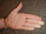
Pallor is a pale color of the skin which can be caused by illness, emotional shock or stress, stimulant use, or anaemia, and is the result of a reduced amount of oxy hemoglobin in skin or mucous membrane.Pallor is more evident on the face ,palms,conjunctiva,tongue etc.. It can develop suddenly or gradually, depending on the cause. It is not usually clinically significant unless it is accompanied by a general pallor pale lips,tongue,palms,mouth etc.
Common causes:
  • anaemia, due to blood loss, poor nutrition, or underlying disease such as sickle cell anaemia 
  • shock, a medical emergency caused by illness or injury 
  • frost bite 
  • underlying cancer 
  • leukemia 
  • peripheral vascular disease 
  • emotional response, due to fear,embarassment ,grief etc. 
  • lead poisoning 
  • reaction to ethanol and/or other drugs such as cannabis, 
  • malaria 
  • hemolytic anemia 
  • congenital heart disease 
  • chronic renal disease 
  • iron/folate/B12 deficiency 
  • G6PD deficiency 
  • GI Bleeding 
  • worm infestations 
  • malnutrition 

Paleness, also known as pale complexion or pallor, is an unusual lightness of skin color when compared with your normal hue. Skin color is determined by several factors, such as the amount of blood flowing to the skin, skin thickness, and the amount of melanin in the skin. Paleness is caused by reduced blood flow or a decreased number of red blood cells. Paleness can be generalized (all over) or local.

Anemia, a condition in which the body doesn’t produce enough red blood cells, is one of the most common causes of paleness. Anaemia can be acute (sudden onset) or chronic (developing slowly).

Acute anemia is usually the result of rapid blood loss from trauma, surgery, bleeding stomach ulcers, or bleeding from the colon.Symptoms of acute onset anemia include:rapid heart rate, shortness of breath,high or low blood pressure,loss of consciousness,irregular or absent menstrual period (DUB) etc.

Chronic anemia is very common. It can be caused by not having enough iron, B12 or folate in your diet. There are also genetic causes of anemia such as sickle cell disease and thalassemia (a genetic disorder that destroys red blood cells). Anemia that develops more slowly can be caused by diseases such as chronic kidney failure or hypothyroidism (when the body does not produce enough thyroid hormone). Certain cancers that affect the bones or bone marrow can also cause anemia due to slow blood loss over a period of weeks to months.

Local paleness usually involves one limb. You should see your doctor if you have sudden onset of generalized paleness or paleness of a limb.Arterial blockage (poor or lack of blood circulation) can cause localized paleness, typically in arms or legs. The limb becomes painful and cold due to lack of circulation.Untreated arterial blockage of a limb can result in gangrene, which can result in the loss of a limb.
Paleness accompanied by signs of blood loss such as fainting, vomiting blood, bleeding from the rectum or abdominal pain is considered a medical emergency. Shortness of breath and sudden onset of paleness, pain and coldness of a limb are also serious symptoms that require immediate medical attention.Your doctor will also review your medical history and perform a physical examination to check your vital signs (heart rate and blood pressure). Pallor can often be diagnosed on sight, but can be hard to detect in people with dark complexions.

Investigations: 
CBC (complete blood count, evaluates if you have anemia),reticulocyte count (a blood test that shows if your bone marrow is replacing blood loss),stool test for the presence of blood,,thyroid function tests (low levels of thyroid hormone causes anemia),BUN and creatinine (kidney function tests),serum iron, B12, and folate levels (to see if nutritional deficiency is causing anemia)
Treatment
  • Balanced diet, and iron (oral/parenteral), B12 or folate supplements,vitamin supplements 
  • Erythropoietin to stimulate RBC production
  • Blood Transfusion may be necessary in some cases 
  • Anti-malarials,de-worming therapy, 
  • surgery is an option for certain causes of acute blood loss, such as trauma. Surgery may also be required for treatment of arterial blockage. 
The long-term consequences for non-treatment of pallor depend upon the underlying cause. Untreated anemia due to blood loss can be fatal. Severe nutritional anemia can lead to other long-term health issues recurrent infections,cardiac failure,.

.


The development of pallor can be acute and associated with a life-threatening illness, or it can be chronic and subtle, occasionally first noted by someone who sees the child less often than the parents. The onset of pallor can provoke anxiety for parents who are familiar with the descriptions of the presentation of leukemia in childhood. In some instances, only reassurance may be needed, as in the case of a light complexioned or fair-skinned, non-anemic child.

Clinically, pallor caused by anemia usually can be appreciated when the hemoglobin concentration is below 8 to 9 g/d.The concentration of hemoglobin in the blood can be lowered by three basic mechanisms:
  • Decreased erythrocyte production
  • Increased erythrocyte destruction
  • Blood loss
WHO's Hemoglobin thresholds used to define anemia(1 g/dL = 0.6206 mmol/L)
Age or gender groupHb threshold (g/dl)Hb threshold (mmol/l)
Children (0.5–5.0 yrs)
11.0
6.8
Children (5–12 yrs)
11.5
7.1
Teens (12–15 yrs)
12.0
7.4
Women, non-pregnant (>15yrs)
12.0
7.4
Women, pregnant
11.0
6.8
Men (>15yrs)
13.0
8.1

Friday, October 18, 2013

EMERGENCIES ALL SHOULD KNOW:ACUTE URINARY RETENTION

ACUTE URINARY RETENTION

 Acute retention causing complete anuria is a medical emergency, as the bladder is filled with urine and can stretch to enormous sizes and possibly tear if not dealt with in time. If the bladder distends more, it becomes painful. 

The increase in bladder pressure can also prevent urine from entering the ureters or even cause urine to pass back up the ureters and get into the kidneys, causing hydronephrosis,sometimes kidney failure, and sepsis. A person should go straight to an emergency department  as soon as possible if unable to urinate for a long time and having a  painfully full bladder.
The causes are
Central causes
  • Consumption of some drugs like amphetamine etc.,
  • patients suffering from mental retardation,multiple sclerosis,stroke,diabetic autonomic neuropathy, 

In the bladder
  • neurogenic bladder,spinal cord diseases,bladder neck contracture,etc.


In the prostate
  • Prostate enlargement either benign or malignant

  urethra
  • Congenital urethral valves
  •  pinhole meatus
  • Obstruction in the urethra, for example a stricture (usually caused either by injury or STD), 
  • obstruction in urethra due to a stone
Urinary retention often occurs without warning. It is basically the inability to pass urine. In some people, the disorder starts gradually but in others it may appear suddenly. Acute urinary retention is a medical emergency and requires prompt treatment. The pain can be excruciating when urine is not able to flow out. Moreover one can develop severe sweating,chest pain,high blood pressure.

In the longer term, obstruction of the urinary tract may cause:
  • Bladder stones
  • weakness of detrusor muscle of bladder (atonic bladder is an extreme form)
  • Hydronephrosis of one/both kidneys leading to renal dysfunction
  • Diverticula (formation of pouches) in the bladder wall (which can lead to urine stasis ,stones and infection)

In acute urinary retention, catheterisation or suprapubic cystostomy (SPC) relieves the retention. These catheters are  inserted by preferrably urologist/trained health care professionals under proper antibiotic cover.

 If the procedure is not done in a sterile fashion, it can introduce infection into the bladder. This can result in an infection of the entire urinary tract. Therefore, sterile technique is a must when inserting a foley catheter. Careful washing of hands, meatus, and reusable catheters are also necessary with clean self catheterization techniques.
In the longer term, treatment depends on the cause.

 BPH may respond to Alpha blocker therapy/laser prostate.
 Some people with BPH are treated with medications like finasteride or dutasteride to decrease prostate enlargement. The drugs only work for mild cases of BPH but also have mild side effects. Some of the medications decrease libido and may cause giddiness,/ fatigue.
Older patients with ongoing problems may require continued intermittent self catheterisation in case of neurogenic bladder.

When you urinate, the brain signals the bladder muscle to tighten, squeezing urine out of the bladder. At the same time, the brain signals the sphincter muscles to relax. As these muscles relax, urine exits the bladder through the urethra. When all the signals occur in the correct order, normal urination occurs.In case of detrusor-sphincter-dyssynergia (DSD) is a condition where some brain/spinal cord diseases contribute to retention of urine.

Urinary retention is a common disorder in elderly males. The most common cause of urinary retention is BPH. This disorder starts around age 50 and symptoms may appear after 10–15 years. BPH is a progressive disorder and narrows the neck of the bladder leading to urinary retention. By the age of 70, almost 10 percent of males have some degree of BPH and 33% have it by the eighth decade of life. While BPH rarely causes sudden urinary retention, the condition can become acute in the presence of certain medications like anti-parkinsonism drugs,anti-psychotics, etc.
In young males, one of the the most common causes of urinary retention is acute infection of the prostate and  the infection is acquired during sexual intercourse.The other common cause is urethral stricture which will be treated succesfully by Optical urethrotomy where the urologist will  incise the stricture under anaesthesia cover.
A woman may experience urinary retention if her bladder sags or moves out of the normal position, a condition called cystocele.The abnormal position of the bladder may cause urine to remain trapped.Cystocele and rectocele are often the results of a dropping of the pelvic support floor for the bladder as seen in mulltiparae,elderly women etc.These are successfully treated by a dedicated urology team.


Wednesday, October 16, 2013

EMERGENCIES ALL SHOULD KNOW:PRIAPISM

PRIAPISM

Priapism is a condition in which the erect penis does not return to its flaccid state, despite the absence of both physical and psychological stimulation, within four hours. Priapism is considered a medical emergency which should receive proper treatment by a qualified medical practitioner. 

The condition develops when blood in the penis becomes trapped and unable to drain. If the condition is not treated immediately, it can lead to scarring and permanent erectile dysfunction.It can occur in all age groups, including newborns.
There are two types of priapism: low-flow and high-flow; 80% to 90% of clinically presented priapisms are low flow disorders. Low-flow involves the blood not adequately returning to the body from the organ. High-flow involves a short-circuit of the vascular system partway along the organ. Treatment is different for each type. Early treatment can be beneficial for a functional recovery.

Priapus is a fertility god (greek)  represented with a disproportionately large and permanent erection.
In the normal erection process, blood flows into the penis and, usually following an orgasm, drains out of the penis without discomfort. When priapism occurs the blood is unable to drain as it would -normally occur.. Because there is little room in the penis for blood to circulate, it becomes stagnant and begins to lose oxygen. Without oxygen, red blood cells become stiff, making proper penis drainage even more problematic.

The causative mechanisms are poorly understood but involve complex neurological and vascular factors. Any bodily ailment that causes blood to thicken or causes red blood cells to lose their flexibility and mobility can lead to priapism. Priapism may be associated with blood disorders, like sickle cell disease, malaria and other conditions such as leukemia, thalassemia, and neurological diseases  such as spinal cord lesions lesions and spinal cord trauma. It has been estimated that approximately 42% of adults with sickle-cell disease will eventually develop priapism.

 Priapism can also be caused by reactions to drugs like desyrel, used to treat depression, or thorazine, used to treat certain mental illnesses ,marijuana and cocaine.The most common medications that cause priapism are intra-cavernous injections for penile doppler,or treatment of erectile dysfunction, like papaverine etc.

Potential complications include ischemia, clotting of the blood retained in the penis (thrombosis), and damage to the blood vessels of the penis which may result in an impaired erectile function or impotence. In serious cases, the ischemia may result in gangrene, which could necessitate penectomy

.Medical advice should be sought immediately for cases of erection beyond four hours and one should be honest in giving proper information about situation/drug intake if any,  leading to priapism.
 Apart from analgesics, locally Ice is applied to the penis and perineum may reduce swelling.

If there is still no relief,the treatment at this stage is to aspirate blood from the corpus cavernosum under sedation with the help of anaesthesiologist.. If this is still insufficient, then intra cavernosal injections of phenylephrine are administered. This should only be performed by a urologist/andrologist/specialist trained in the procedure, with the patient under constant ECG monitoring, as phenylephrine can cause severe hypertension, bradycardia/tachycardia,arrhythmia 
.As the complication of shortened, indurated and non-erectile penis is high in prolonged priapism, early penile prosthesis implantation can be performed. Apart from early resumption of sexual activity, early implantation can avoid the formation of dense fibrosis and hence a shortened penis.

A general rule of thumb is to pay close attention to excess swelling or pain in the penis and to seek out care sooner rather than later if you suspect something ABNORMAL.

Tuesday, October 15, 2013

EMERGENCIES ALL SHOULD KNOW:RENAL COLIC

RENAL COLIC


The renal colic is a pain related to stones in urinary tract, comes in waves due to ureteric peristalsis. It may come in two varieties: dull and acute; the acute variation is particularly unpleasant and is often described as one of the strongest pain sensations felt by humans (being worse than childbirth, broken bones, gunshot wounds, burns, or surgery).

Passing kidney stones can be quite painful, the experience is said to be traumatizing due to pain, and the experience of passing blood, blood clots, and pieces of the stone. The majority of renal calculi contain calcium. The pain generated by renal colic is primarily caused by dilation, stretching, and spasm because of the acute ureteral obstruction.

Depending on the sufferer's situation, surgery may be needed to remove the stone which is impacted in the  urinary passage anywhere between kidney and urethra in both males and females.One may have kidney stones in one or both  kidneys or ureters. Blood clots, ureter spasms (tightening and relaxing), and dead tissue may also block the urinary tract.The pain may start quickly, come and go, and may become worse over time. One may have any of the following:

  • Severe low back, abdominal, or groin pain. The pain may be so bad that you are not able to sit still. You may have pain when you urinate. The pain may also cause you to sweat and feel like your heart is beating faster than usual.
  • Nausea and vomiting.
  • Feeling the need to urinate often, or right away.
  • Urinating less than what is normal for you, or not at all.
Most small stones are passed spontaneously and only pain killers and anti-spasmodics are required.There is typically no antalgic position for the patient (lying down on the non-aching side etc).


Medical treatment of nephrolithiasis involves supportive care and administration of agents, such as the following:
  • IV hydration
  • IV narcotic analgesics
  • NSAIDS 
  • Uricosuric agents (eg, allopurinol)
  • Antiemetics
  • Antibiotics 
  • Alkalinizing agents (eg, potassium citrate, sodium bicarbonate): For uric acid and cysteine calculi
  • Alpha blockers (eg, tamsulosin, terazosin)


    The reason why kidney stones are formed is still not known in many cases. Many waste chemicals are dissolved in the urine. The chemicals sometimes form tiny crystals in the concentrated urine which clump together to form a small stone.About half of the people who have a kidney stone develop another one at a later age in life.

    A stone that is stuck in a kidney may cause pain in the side of the abdomen.This is a severe pain which comes and goes and is caused by a stone that passes into the ureter (the tube that leads from the kidney to the bladder). The stone becomes stuck. The ureter squeezes the stone towards the bladder, which causes intense pain in the side of the abdomen. The pain may spread down into the lower abdomen or groin. The patient  may sweat or feel sick due to the pain.One may also see blood in  urine (urine turns red) caused by a stone rubbing against the inside of the ureter.Urine infections are more common in people with kidney stones. Infections can cause fever, pain on passing urine and increased frequency of passing urine.
 One is  more likely to form a stone if urine is concentrated. For example, if you exercise vigorously, if you live in a hot climate or if you work in a hot environment when you may lose more fluid as sweat and less as urine.
You are also more prone to develop kidney stones if you have:
  • Recurrent urine or kidney infections.
  • A kidney with scars or cysts on it.
  • A close relative who has had a kidney stone.
The following are the battery of tests done if a person is having renal colic:
  • Blood tests: routine blood tests,serum urea,creatinine  to know the function of kidneys,etc.
  • Urine sample: A sample of your urine is collected and sent to a lab for tests for any infections.
  • Renal ultrasound: A renal ultrasound is a test using sound waves to look at your kidneys. An ultrasound may show if you have a kidney stone or other problems that are causing your pain.
    • Computed tomography scan: A computed tomography (CT) scan is a special x-ray using a computer to take pictures of your urinary tract. A CT scan may be done to check for stones and their size. A CT scan may also be done to check for other causes of your pain.
    • KUB x-ray: A KUB  x-ray is a picture of your kidneys (K), ureters (U), and bladder (B).
    • Intravenous pyelogram: An intravenous pyelogram (IVP) is an x-ray of your kidneys, ureters, and bladder. Dye is put into your IV before the pictures are taken.  People who are allergic to shellfish may be allergic to some dyes. You may need to have more than one x-ray over short periods of time during your IVP.
  •    An analysis of the stone if you pass it out. To catch a stone, pass urine through gauze, a tea strainer or a filter    such as a coffee filter.
  • The location and characteristics of pain in is related to site of stone in urinary tract:
    • Stones obstructing ureteropelvic junction: Mild to severe deep flank pain without radiation to the groin; irritative voiding symptoms (eg, frequency, dysuria); suprapubic pain, urinary frequency/urgency, dysuria, stranguria, bowel symptoms
    • Stones within ureter: Abrupt, severe, colicky pain in the flank and ipsilateral lower abdomen; radiation to testicles or vulvar area; intense nausea with or without vomiting
    • Upper ureteral stones: Radiate to flank or lumbar areas
    • Midureteral calculi: Radiate anteriorly and caudally
    • Distal ureteral stones: Radiate into groin or testicle (men) or labia majora (women)
    • Stones passed into bladder: Mostly asymptomatic; rarely, positional urinary retention
  • Management:
  • Some stones that form and stick in the kidney do not cause symptoms or any harm. They can just be left if they are small. Sometimes you may be offered medicines to help the small stone pass through, such as  tamsulosin.
  • The size of the stone is an important predictor of spontaneous passage. A stone less than 4 mm in diameter has an 80% chance of spontaneous passage; this falls to 20% for stones larger than 8 mm in diameter. However, stone passage also depends on the exact shape and location of the stone and the specific anatomy of the upper urinary tract in the particular individual.
       
Some stones become stuck in a ureter or kidney and cause persistent symptoms or problems. In these cases, the pain usually becomes severe and you may need to be admitted to hospital. There are various treatment options which include the following:


  • Ureteroscopy is another treatment that may be used. In this procedure, a thin telescope either (flexible or rigid) is passed up into the ureter via the urethra and bladder. Once the stone is seen, a laser (or other form of energy) is used to break up the stone. This technique is suitable for most types of stone. The stone removal is followed by the  placement of DJ(Internal ureteral) stents which form a coil at either end, One coil forms in the renal pelvis and the other in the bladder. Stents are available in lengths from 20-30 cm and in 3 widths from 4.6F to 8.5F. The stents allow proper functioning of kidneys and are later removed usually after 4 weeks by a minor procedure.
  • Extracorporeal shock wave lithotripsy (ESWL). This uses high-energy shock waves which are focused on to the stones from a machine outside the body to break up stones. You then pass out the tiny broken fragments when you pass urine.

  • Percutaneous nephrolithotomy (PCNL) is used for some select stones not suitable for ESWL. A nephroscope (a thin telescope-like instrument) is passed through the skin and into the kidney. The stone is broken up and the fragments of stone are removed via the nephroscope. This procedure is usually done under general anaesthetic.
About half of people who have a kidney stone develop another one within 10 years. Sometimes stones can be prevented from forming.f you have had one stone, you are less likely to have a recurrence if you drink plenty of fluid, mainly water, throughout the day (and night). The aim is to keep the urine dilute. (Your urine is more dilute if it is clear of colour rather than a dark yellow colour.) To do this, you should drink between two and three litres a day (unless your doctor advises otherwise if you have other medical problems). If you work or live in a hot environment, you should drink even more.


  • A dietitian may advise people with calcium oxalate stones to reduce the oxalate content of their diet. This may include reducing rhubarb, coffee and spinach.
  • Uric acid stones can be prevented with a medicine.
more in blog
PRIAPISM
ACUTE URINE RETENTION
SEVERE PALLOR
ABSCESS
ANIMAL BITES
POISONING
BURNS
SEIZURES
PARALYTIC STROKE
ANY PREGNANCY RELATED EVENT
SUDDEN LOSS OR IMPAIRMENT OF VISION
SUDDEN HEAD ACHE AND VOMITING
HIGH GRADE FEVER
NON-RESPONSIVENESS
VIOLENT BEHAVIOUR
SUDDEN CALF PAIN


SEVERE BREATHLESSNESS