Pre-operative ultrasound (Before first surgery) showing cystic mass
The laparoscopy was abandoned because contrary to their expectations they found retroperitoneal mass on the right side of the retroperitoneum.
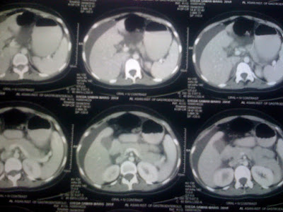
She came to us with a post-operative contrast enhanced CT Scan which revealed urinoma near middle of the right ureter and diffuse ascites. The urinoma was encapsulated in thick capsule (? Chronic process).


On clinical examination she was looking ill and frail. She had loose motions(? Pelvic collection induced).
Her vitals though were maintained except for tachycardia.She had normal hematological and biochemical parameters. Her abdomen was mildly distended with urinary leakage through one of the ports.
She was taken up for Retrograde Pyelography which showed mid-ureteric disruption and dye leaking into a diffuse cavity.The patient was made prone for percutaneous nephrostomy drainage.
Right Percutaneous Drainage was performed through a midcalyceal approach for possible antegrade stenting sometimes in future.
She started draining around 100 ml of urine per hour through the nephrostomy and her leakage of urine through the port and the abdominal distension subsided.Her loose motions also subsided
The very next day she started looking fresh and was back to her normal routine.
She is planned to undergo an evaluation after a period of 6 weeks hoping that till that time the urinoma would subside and the inflammatory reaction would also subside.Then a definitive plan for ureteric reconstruction will be taken up.
This is a rare case of spontaneous urinoma at level of mid-ureter with no apparent aetiology clinically,history and imaging wise.
Having searched the English Literature the urinoma spontaneous in this location was found to be very rare --due to abdominal aortic aneurysm or retroperitoneal fibrosis.In this case both the factors were not seen on CT scan.The possibility of tuberculous lymphadenopathy involving the ureter causing ischemic necrosis of that particular segment leading to spontaneous urinoma is being kept in mind.
She will be investigated for quantiferon TB TEST in the interim .With all said and done probably exploration after 6 weeks and biopsy of the region only might give a definitive clue.


No comments:
Post a Comment